Department of Orthopedic Surgery, University of California at Los Angeles, Rehabilitation Building 22-69, 1000 Veteran Avenue, Los Angeles, CA 90095, USA.
J Theor Biol. 2011 Sep 21;285(1):147-55. doi: 10.1016/j.jtbi.2011.06.016. Epub 2011 Jun 23.
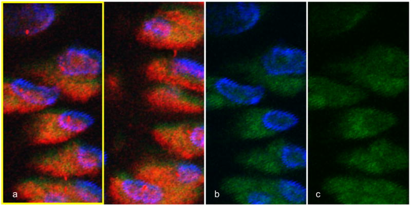
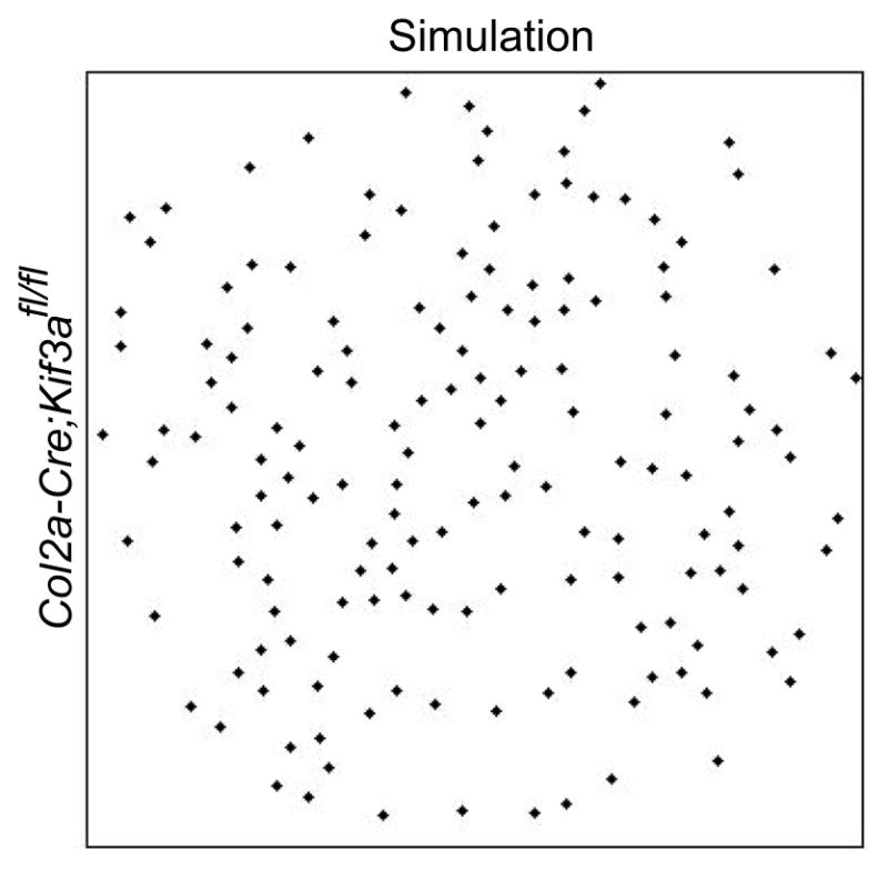
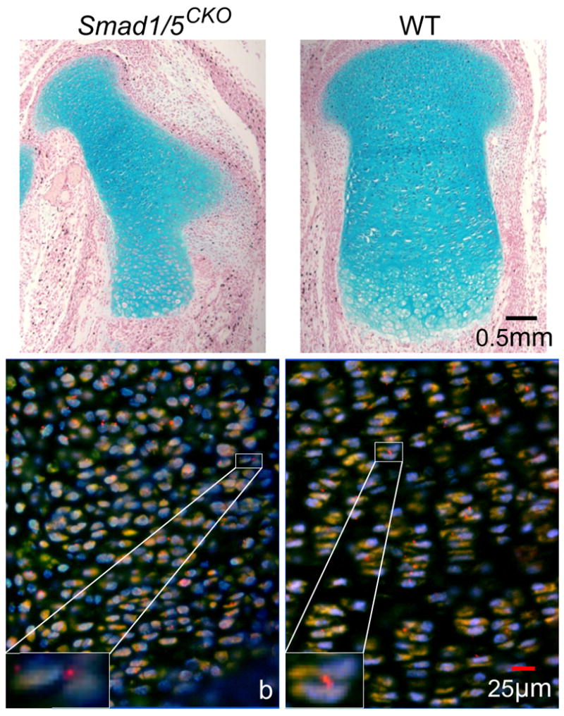
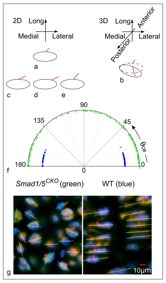
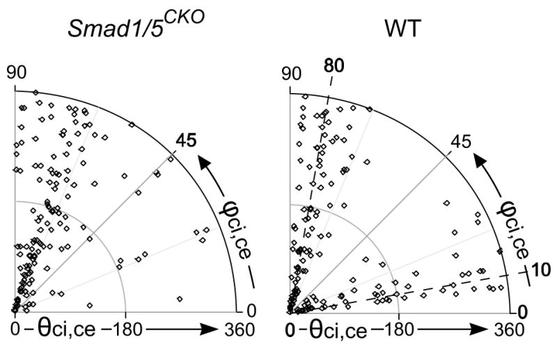
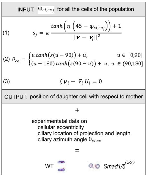
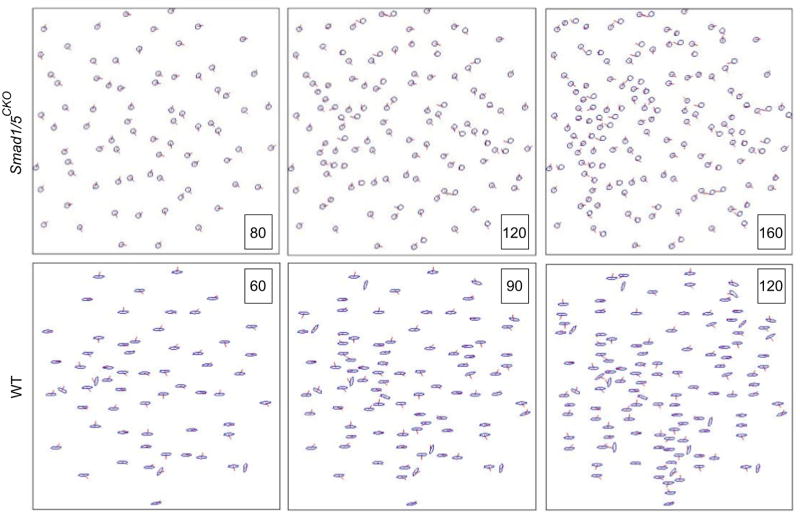
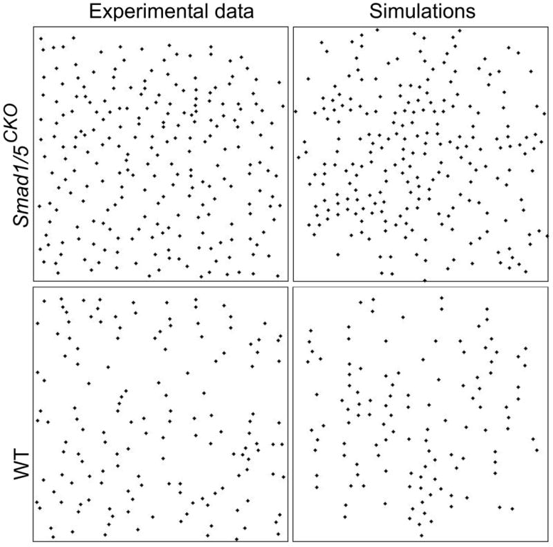
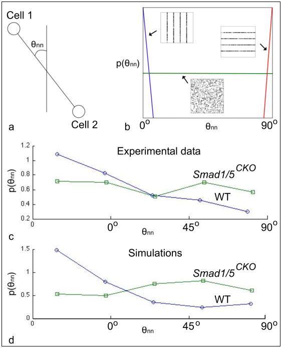
The research investigates the role of the immotile chondrocytic primary cilium in the growth plate. This study was motivated by (i) the recent evidence of the mechano-sensorial function of the primary cilium in kidney tubule epithelial cells and (ii) the distinct three-dimensional orientation patterns that the chondrocytic primary cilium forms in articular cartilage in the presence or the absence of loading. For our investigation, we used the Smad1/5(CKO) mutant mouse, whose disorganized growth plate is due to the conditional deletion of Smad 1 and 5 proteins that also affect the so-called Indian Hedgehog pathway, whose physical and functional topography has been shown to be partially controlled by the primary cilium. Fluorescence and confocal microscopy on stained sections visualized ciliated chondrocytes. Morphometric data regarding position, orientation and eccentricity of chondrocytes, and ciliary localization on cell membrane, length and orientation, were collected and reconstructed from images. We established that both localization and orientation of the cilium are definite, and differently so, in the Smad1/5(CKO) and control mice. The orientation of the primary cilium, relative to the major axis of the chondrocyte, clusters at 80° with respect to the anterior-posterior direction for the Smad1/5(CKO) mice, showing loss of the additional clustering present in the control mice at 10°. We therefore hypothesized that the clustering at 10° contains information of columnar organization. To test our hypothesis, we prepared a mathematical model of relative positioning of the proliferative chondrocytic population based on ciliary orientation. Our model belongs to the category of "interactive particle system models for self-organization with birth". The model qualitatively reproduced the experimentally observed chondrocytic arrangements in growth plate of each of the Smad1/5(CKO) and control mice. Our mathematically predicted cell division process will need to be observed experimentally to advance the identification of ciliary function in the growth plate.
本研究旨在探讨静息状态下软骨细胞初级纤毛在生长板中的作用。该研究的动机有二:(i)最近有证据表明,初级纤毛在肾小管上皮细胞中具有机械感受功能;(ii)在存在或不存在载荷的情况下,软骨细胞初级纤毛在关节软骨中形成的独特三维定向模式。在我们的研究中,我们使用了 Smad1/5(CKO) 突变小鼠,其生长板紊乱是由于 Smad 1 和 5 蛋白的条件性缺失所致,这些蛋白也影响所谓的印度刺猬途径,其物理和功能拓扑结构已被证明部分受初级纤毛控制。染色切片的荧光和共聚焦显微镜可观察到纤毛软骨细胞。关于软骨细胞的位置、定向和偏心率,以及质膜上的纤毛定位、长度和定向的形态计量数据,是从图像中收集和重建的。我们确定,Smad1/5(CKO)和对照小鼠中,纤毛的定位和定向都是确定的,而且方式不同。相对于软骨细胞的长轴,初级纤毛的定向在 Smad1/5(CKO)小鼠中以 80°聚类,相对于前-后方向,显示出对照小鼠中存在的额外聚类缺失,在 10°聚类。因此,我们假设 10°聚类包含柱状组织的信息。为了验证我们的假设,我们根据纤毛定向制备了一个基于增殖性软骨细胞群体相对定位的数学模型。我们的模型属于“具有出生的自组织的交互式粒子系统模型”类别。该模型定性地再现了 Smad1/5(CKO)和对照小鼠生长板中观察到的软骨细胞排列。我们预测的细胞分裂过程需要通过实验观察来推进对生长板中纤毛功能的识别。