Anantham Devanand, Phua Ghee-Chee, Low Su-Ying, Koh Mariko-Siyue
Department of Respiratory and Critical Care Medicine, Singapore General Hospital, Outram Road, Singapore 169608.
Diagn Ther Endosc. 2011;2011:468237. doi: 10.1155/2011/468237. Epub 2011 May 11.
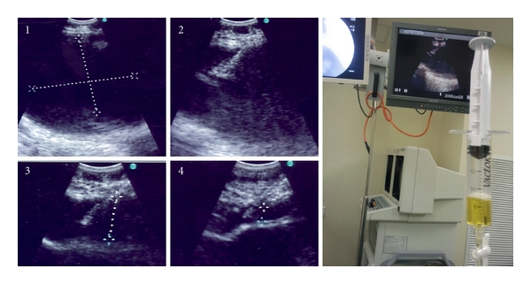
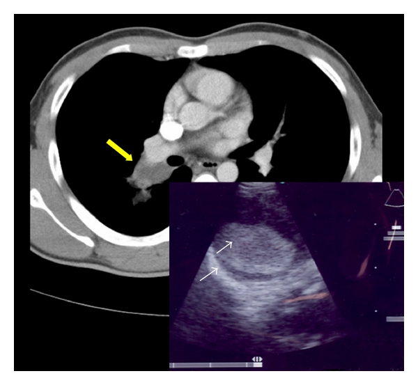
Diagnosis of bronchogenic cysts is possible with computed tomography, but half of all cases present as soft tissue densities. Two such cases are highlighted where asymptomatic bronchogenic cysts that presented as soft tissue masses were evaluated by endobronchial ultrasound (EBUS). After studying the ultrasound image characteristics, the diagnosis was confirmed using EBUS-guided transbronchial needle aspiration (EBUS-TBNA). The first case had ultrasound findings of an anechoic collection, and the aspirate was serous with negative microbiologic cultures. The second was an echogenic collection within a hyperechoic wall. Needle aspirate was purulent and cultured Haemophilus influenza. The diagnosis of a bronchogenic cyst complicated by infection was made, and the lesion was surgically resected. This potential for EBUS in the diagnosis of bronchogenic cysts and in identifying complications such as infection should be considered in the management of such cases.
通过计算机断层扫描可以诊断支气管囊肿,但所有病例中有一半表现为软组织密度。本文重点介绍了两例通过支气管内超声(EBUS)评估的无症状支气管囊肿病例,这些囊肿表现为软组织肿块。在研究超声图像特征后,通过EBUS引导的经支气管针吸活检(EBUS-TBNA)确诊。第一例超声表现为无回声液性暗区,吸出液为浆液性,微生物培养阴性。第二例为高回声壁内的回声增强区。针吸物为脓性,培养出流感嗜血杆菌。诊断为合并感染的支气管囊肿,病变行手术切除。在这类病例的管理中,应考虑EBUS在支气管囊肿诊断以及识别感染等并发症方面的潜力。