Bassoli S, Borsari S, Ferrari C, Giusti F, Pellacani G, Ponti G, Seidenari S
Department of Dermatology, University of Modena and Reggio Emilia, Modena, Italy.
J Skin Cancer. 2011;2011:180980. doi: 10.1155/2011/180980. Epub 2011 Mar 23.
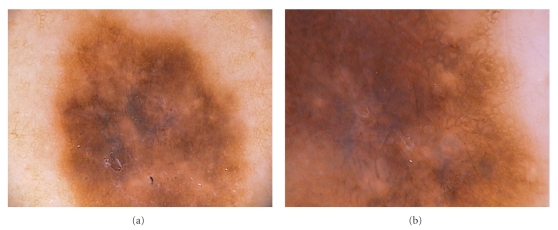
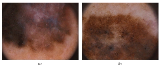
As fibrosis and melanosis are often seen in malignant melanoma, the presence of dermoscopic signs of regression may represent a clue for the diagnosis of malignancy. Our aim was to assess the frequency and extent of 11 dermoscopic features of regression evaluating dermoscopic images of 111 melanomas in situ (MIS). Regression structures (grey-blue areas, white areas, peppering, and/or blue-whitish veil) were present in 80.1% of the lesions. Approximately 80% of the lesions showed regression of dermoscopic structures and light brown areas. Most lesions showed the presence of grey-blue areas (74.7%), whereas peppering was observable in 30.6% of MIS. Areas of fibrosis were mainly observable as structureless areas with a pinkish hue (50.4%). Based on our data, the reticular pattern of blue regression and light brown areas can be considered a significant discriminator and a reliable predictor of MIS.
由于纤维化和黑变病在恶性黑色素瘤中较为常见,皮肤镜下消退迹象的存在可能是诊断恶性肿瘤的线索。我们的目的是评估11种消退皮肤镜特征的出现频率和范围,为此对111例原位黑色素瘤(MIS)的皮肤镜图像进行了评估。80.1%的病变存在消退结构(灰蓝色区域、白色区域、点状分布和/或蓝白色薄纱)。约80%的病变显示皮肤镜结构和浅棕色区域出现消退。大多数病变存在灰蓝色区域(74.7%),而30.6%的原位黑色素瘤可见点状分布。纤维化区域主要表现为无结构的淡粉色区域(50.4%)。根据我们的数据,蓝色消退的网状模式和浅棕色区域可被视为原位黑色素瘤的重要鉴别特征和可靠预测指标。