• 文献检索
  • 文档翻译
  • 深度研究
  • 学术资讯
  • Suppr Zotero 插件Zotero 插件
  • 邀请有礼
  • 套餐&价格
  • 历史记录
应用&插件
Suppr Zotero 插件Zotero 插件浏览器插件Mac 客户端Windows 客户端微信小程序
定价
高级版会员购买积分包购买API积分包
服务
文献检索文档翻译深度研究API 文档MCP 服务
关于我们
关于 Suppr公司介绍联系我们用户协议隐私条款
关注我们

Suppr 超能文献

核心技术专利:CN118964589B侵权必究
粤ICP备2023148730 号-1Suppr @ 2026

文献检索

告别复杂PubMed语法,用中文像聊天一样搜索,搜遍4000万医学文献。AI智能推荐,让科研检索更轻松。

立即免费搜索

文件翻译

保留排版,准确专业,支持PDF/Word/PPT等文件格式,支持 12+语言互译。

免费翻译文档

深度研究

AI帮你快速写综述,25分钟生成高质量综述,智能提取关键信息,辅助科研写作。

立即免费体验

多重酶谱法可捕捉到组织蛋白酶 K、L 和 S 在人乳腺癌、肺癌和宫颈癌中的特定阶段的活性谱。

Multiplex zymography captures stage-specific activity profiles of cathepsins K, L, and S in human breast, lung, and cervical cancer.

机构信息

Wallace H. Coulter Department of Biomedical Engineering, Georgia Institute of Technology and Emory University, GA 30332, Atlanta, USA.

出版信息

J Transl Med. 2011 Jul 14;9:109. doi: 10.1186/1479-5876-9-109.

DOI:10.1186/1479-5876-9-109
PMID:21756348
原文链接:https://pmc.ncbi.nlm.nih.gov/articles/PMC3146840/
Abstract

BACKGROUND

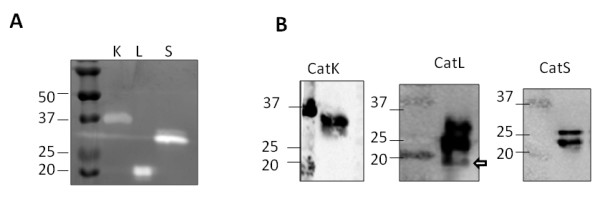
Cathepsins K, L, and S are cysteine proteases upregulated in cancer and proteolyze extracellular matrix to facilitate metastasis, but difficulty distinguishing specific cathepsin activity in complex tissue extracts confounds scientific studies and employing them for use in clinical diagnoses. Here, we have developed multiplex cathepsin zymography to profile cathepsins K, L, and S activity in 10 μg human breast, lung, and cervical tumors by exploiting unique electrophoretic mobility and renaturation properties.

METHODS

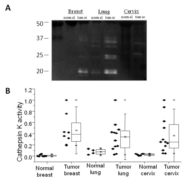
Frozen breast, lung, and cervix cancer tissue lysates and normal organ tissue lysates from the same human patients were obtained (28 breast tissues, 23 lung tissues, and 23 cervix tissues), minced and homogenized prior to loading for cathepsin gelatin zymography to determine enzymatic activity.

RESULTS

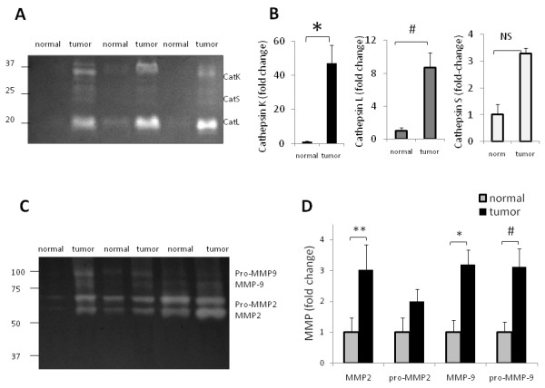
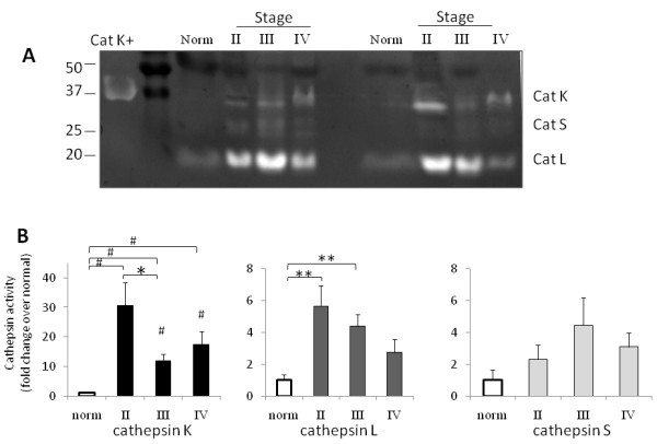
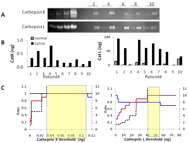
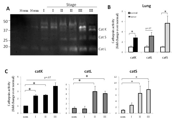
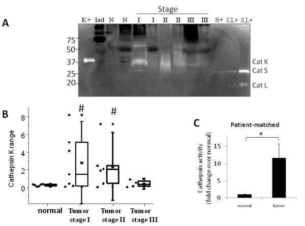
Cleared bands of cathepsin activity were identified and validated in tumor extracts and detected organ- and stage-specific differences in activity. Cathepsin K was unique compared to cathepsins L and S. It was significantly higher for all cancers even at the earliest stage tested (stage I for lung and cervix (n = 6, p < .05), and stage II for breast; n = 6, p < .0001). Interestingly, cervical and breast tumor cathepsin activity was highest at the earliest stage we tested, stages I and II, respectively, and then were significantly lower at the latest stages tested (III and IV, respectively) (n = 6, p < 0.01 and p < 0.05), but lung cathepsin activity increased from one stage to the next (n = 6, p < .05). Using cathepsin K as a diagnostic biomarker for breast cancer detected with multiplex zymography, yielded 100% sensitivity and specificity for 20 breast tissue samples tested (10 normal; 10 tumor) in part due to the consistent absence of cathepsin K in normal breast tissue across all patients.

CONCLUSIONS

To summarize, this sensitive assay provides quantitative outputs of cathepsins K, L, and S activities from mere micrograms of tissue and has potential use as a supplement to histological methods of clinical diagnoses of biopsied human tissue.

摘要

背景

组织蛋白酶 K、L 和 S 是在癌症中上调的半胱氨酸蛋白酶,可降解细胞外基质以促进转移,但在复杂组织提取物中难以区分特定的组织蛋白酶活性,这给科学研究带来了困难,并阻碍了它们在临床诊断中的应用。在这里,我们开发了多重组织蛋白酶酶谱法,通过利用独特的电泳迁移率和复性特性,在 10μg 人乳腺、肺和宫颈肿瘤中对组织蛋白酶 K、L 和 S 的活性进行分析。

方法

获取冷冻的乳腺、肺和宫颈癌组织和来自同一患者的正常器官组织的组织匀浆(28 份乳腺组织、23 份肺组织和 23 份宫颈组织),在加载用于组织蛋白酶明胶酶谱法以确定酶活性之前,将其切碎并均化。

结果

在肿瘤提取物中鉴定和验证了清除的组织蛋白酶活性带,并检测到活性的器官和阶段特异性差异。组织蛋白酶 K 与组织蛋白酶 L 和 S 不同。即使在测试的最早阶段(肺和宫颈的 I 期(n=6,p<0.05)和乳腺的 II 期;n=6,p<0.0001),所有癌症的组织蛋白酶 K 水平均显著升高。有趣的是,在我们测试的最早阶段,宫颈和乳腺肿瘤的组织蛋白酶活性最高,分别为 I 期和 II 期,然后在测试的最新阶段(分别为 III 期和 IV 期)显著降低(n=6,p<0.01 和 p<0.05),但肺组织蛋白酶活性从一个阶段增加到下一个阶段(n=6,p<0.05)。使用多重酶谱法作为乳腺癌的诊断生物标志物,检测 20 个组织样本的结果显示 100%的敏感性和特异性(10 个正常;10 个肿瘤),部分原因是在所有患者中,正常乳腺组织中始终不存在组织蛋白酶 K。

结论

总之,这种灵敏的检测方法可以从微量组织中定量输出组织蛋白酶 K、L 和 S 的活性,并有可能作为组织活检的临床诊断中组织学方法的补充。

https://cdn.ncbi.nlm.nih.gov/pmc/blobs/b79e/3146840/4edab67a64b0/1479-5876-9-109-7.jpg
https://cdn.ncbi.nlm.nih.gov/pmc/blobs/b79e/3146840/a63f2e1fe274/1479-5876-9-109-1.jpg
https://cdn.ncbi.nlm.nih.gov/pmc/blobs/b79e/3146840/18765fabe14a/1479-5876-9-109-2.jpg
https://cdn.ncbi.nlm.nih.gov/pmc/blobs/b79e/3146840/bc0edcd327e8/1479-5876-9-109-3.jpg
https://cdn.ncbi.nlm.nih.gov/pmc/blobs/b79e/3146840/4d1ff4ee607f/1479-5876-9-109-4.jpg
https://cdn.ncbi.nlm.nih.gov/pmc/blobs/b79e/3146840/beccaa9d4d64/1479-5876-9-109-5.jpg
https://cdn.ncbi.nlm.nih.gov/pmc/blobs/b79e/3146840/cc64b6750692/1479-5876-9-109-6.jpg
https://cdn.ncbi.nlm.nih.gov/pmc/blobs/b79e/3146840/4edab67a64b0/1479-5876-9-109-7.jpg

相似文献

1
Multiplex zymography captures stage-specific activity profiles of cathepsins K, L, and S in human breast, lung, and cervical cancer.多重酶谱法可捕捉到组织蛋白酶 K、L 和 S 在人乳腺癌、肺癌和宫颈癌中的特定阶段的活性谱。
J Transl Med. 2011 Jul 14;9:109. doi: 10.1186/1479-5876-9-109.
2
Multiplex Cathepsin Zymography to Detect Amounts of Active Cathepsins K, L, S, and V.多重组织蛋白酶酶谱法检测组织蛋白酶K、L、S和V的活性量
Methods Mol Biol. 2017;1626:239-252. doi: 10.1007/978-1-4939-7111-4_23.
3
Manipulating substrate and pH in zymography protocols selectively distinguishes cathepsins K, L, S, and V activity in cells and tissues.在酶谱法方案中对基质和 pH 值进行操作,可以有选择性地区分细胞和组织中组织蛋白酶 K、L、S 和 V 的活性。
Arch Biochem Biophys. 2011 Dec 1;516(1):52-7. doi: 10.1016/j.abb.2011.09.009. Epub 2011 Sep 29.
4
Systematic optimization of multiplex zymography protocol to detect active cathepsins K, L, S, and V in healthy and diseased tissue: compromise among limits of detection, reduced time, and resources.系统优化多重酶谱法检测健康和患病组织中活性组织蛋白酶 K、L、S 和 V 的方案:检测限、缩短时间和资源之间的折衷。
Mol Biotechnol. 2013 Jul;54(3):1038-47. doi: 10.1007/s12033-013-9658-4.
5
Differential cathepsin responses to inhibitor-induced feedback: E-64 and cystatin C elevate active cathepsin S and suppress active cathepsin L in breast cancer cells.组织蛋白酶对抑制剂诱导反馈的差异反应:E-64和胱抑素C提高乳腺癌细胞中组织蛋白酶S的活性并抑制组织蛋白酶L的活性。
Int J Biochem Cell Biol. 2016 Oct;79:199-208. doi: 10.1016/j.biocel.2016.08.030. Epub 2016 Aug 31.
6
Identification and discrimination of extracellularly active cathepsins B and L in high-invasive melanoma cells.高侵袭性黑色素瘤细胞中细胞外活性组织蛋白酶B和L的鉴定与区分
Anal Biochem. 2006 Jun 1;353(1):57-62. doi: 10.1016/j.ab.2006.01.037. Epub 2006 Feb 9.
7
Cathepsins B, H and L in human breast carcinoma.人乳腺癌中的组织蛋白酶B、H和L
Eur J Clin Chem Clin Biochem. 1992 Feb;30(2):69-74.
8
Cathepsins D, B and L in breast carcinoma and in transformed human breast epithelial cells (HBEC).乳腺癌及转化的人乳腺上皮细胞(HBEC)中的组织蛋白酶D、B和L
Biol Chem Hoppe Seyler. 1995 Jun;376(6):357-63. doi: 10.1515/bchm3.1995.376.6.357.
9
The effect of catalase on migration and invasion of lung cancer cells by regulating the activities of cathepsin S, L, and K.过氧化氢酶通过调节组织蛋白酶 S、L 和 K 的活性对肺癌细胞迁移和侵袭的影响。
Exp Cell Res. 2014 Apr 15;323(1):28-40. doi: 10.1016/j.yexcr.2014.02.014. Epub 2014 Feb 26.
10
Simultaneous expression of Cathepsins B and K in pulmonary adenocarcinomas and squamous cell carcinomas predicts poor recurrence-free and overall survival.组织蛋白酶B和K在肺腺癌和肺鳞状细胞癌中的同时表达预示着无复发生存期和总生存期较差。
Lung Cancer. 2009 Apr;64(1):79-85. doi: 10.1016/j.lungcan.2008.07.005. Epub 2008 Aug 29.

引用本文的文献

1
Immunohistochemical Evaluation of Cathepsin B, L, and S Expression in Breast Cancer Patients.乳腺癌患者组织蛋白酶B、L和S表达的免疫组织化学评估
Mol Imaging Biol. 2024 Dec;26(6):1057-1067. doi: 10.1007/s11307-024-01955-5. Epub 2024 Sep 27.
2
Challenges and Opportunities Modeling the Dynamic Tumor Matrisome.动态肿瘤基质组建模的挑战与机遇
BME Front. 2023 Jan 16;4:0006. doi: 10.34133/bmef.0006. eCollection 2023.
3
Mechanical strain modulates extracellular matrix degradation and byproducts in an isoform-specific manner.

本文引用的文献

1
Antibody targeting of cathepsin S inhibits angiogenesis and synergistically enhances anti-VEGF.抗体靶向组织蛋白酶 S 抑制血管生成,并与抗 VEGF 药物协同增强作用。
PLoS One. 2010 Sep 2;5(9):e12543. doi: 10.1371/journal.pone.0012543.
2
Detection of femtomole quantities of mature cathepsin K with zymography.用酶谱法检测成熟组织蛋白酶 K 的皮摩尔量。
Anal Biochem. 2010 Jun 1;401(1):91-8. doi: 10.1016/j.ab.2010.02.035. Epub 2010 Mar 3.
3
The American Joint Committee on Cancer: the 7th edition of the AJCC cancer staging manual and the future of TNM.
机械应变以同种型特异性的方式调节细胞外基质的降解及其产物。
Biochim Biophys Acta Gen Subj. 2023 Mar;1867(3):130286. doi: 10.1016/j.bbagen.2022.130286. Epub 2022 Dec 1.
4
Cysteine Cathepsins in Breast Cancer: Promising Targets for Fluorescence-Guided Surgery.半胱氨酸组织蛋白酶在乳腺癌中的作用:荧光引导手术的有前途的靶点。
Mol Imaging Biol. 2023 Feb;25(1):58-73. doi: 10.1007/s11307-022-01768-4. Epub 2022 Aug 24.
5
Cysteine Cathepsins and Matrix Metalloproteases Among Breast Cancer Patients.乳腺癌患者中的半胱氨酸组织蛋白酶和基质金属蛋白酶
Breast Cancer (Dove Med Press). 2021 May 7;13:271-283. doi: 10.2147/BCTT.S305387. eCollection 2021.
6
A Porcine Organ-Culture Glaucoma Model Mimicking Trabecular Meshwork Damage Using Oxidative Stress.一种利用氧化应激模拟小梁网损伤的猪器官培养青光眼模型
Invest Ophthalmol Vis Sci. 2021 Mar 1;62(3):18. doi: 10.1167/iovs.62.3.18.
7
Cysteine cathepsins are altered by flow within an engineered microvascular niche.半胱氨酸组织蛋白酶在工程化微血管微环境中会因流动而发生改变。
APL Bioeng. 2020 Nov 4;4(4):046102. doi: 10.1063/5.0023342. eCollection 2020 Dec.
8
Cathepsin K regulates localization and secretion of Tartrate-Resistant Acid Phosphatase (TRAP) in TRAP-overexpressing MDA-MB-231 breast cancer cells.组织蛋白酶 K 调节 TRAP 过表达 MDA-MB-231 乳腺癌细胞中 Tartrate-Resistant Acid Phosphatase(TRAP)的定位和分泌。
BMC Mol Cell Biol. 2020 Mar 18;21(1):15. doi: 10.1186/s12860-020-00253-6.
9
Potential role of cytotoxic T-lymphocyte antigen 2 alpha in secretory activity of endocrine cells in mouse adenohypophysis.细胞毒性T淋巴细胞抗原2α在小鼠腺垂体内分泌细胞分泌活动中的潜在作用。
Open Vet J. 2019 Jul;9(2):114-119. doi: 10.4314/ovj.v9i2.4. Epub 2019 Apr 12.
10
CT Imaging of Enzymatic Activity in Cancer Using Covalent Probes Reveal a Size-Dependent Pattern.使用共价探针的癌症酶活性的 CT 成像揭示了一种尺寸依赖性模式。
J Am Chem Soc. 2018 Sep 26;140(38):12010-12020. doi: 10.1021/jacs.8b05817. Epub 2018 Sep 18.
美国癌症联合委员会:第 7 版 AJCC 癌症分期手册与 TNM 的未来。
Ann Surg Oncol. 2010 Jun;17(6):1471-4. doi: 10.1245/s10434-010-0985-4.
4
IL-4 induces cathepsin protease activity in tumor-associated macrophages to promote cancer growth and invasion.IL-4 诱导肿瘤相关巨噬细胞中的组织蛋白酶蛋白酶活性,从而促进癌症生长和侵袭。
Genes Dev. 2010 Feb 1;24(3):241-55. doi: 10.1101/gad.1874010. Epub 2010 Jan 15.
5
Bone marrow-derived cathepsin K cleaves SPARC in bone metastasis.骨髓来源的组织蛋白酶K在骨转移中裂解富含半胱氨酸的酸性分泌蛋白(SPARC)。
Am J Pathol. 2009 Sep;175(3):1255-69. doi: 10.2353/ajpath.2009.080906. Epub 2009 Aug 21.
6
Pharmacological inhibitors to identify roles of cathepsin K in cell-based studies: a comparison of available tools.用于在细胞研究中确定组织蛋白酶K作用的药理学抑制剂:现有工具的比较
Biol Chem. 2009 Sep;390(9):941-8. doi: 10.1515/BC.2009.092.
7
MT1-MMP and type II collagen specify skeletal stem cells and their bone and cartilage progeny.MT1-MMP 和 II 型胶原特异性标记骨骼干细胞及其骨和软骨祖细胞。
J Bone Miner Res. 2009 Nov;24(11):1905-16. doi: 10.1359/jbmr.090510.
8
Usefulness of serum cathepsin L as an independent biomarker in patients with coronary heart disease.血清组织蛋白酶L作为冠心病患者独立生物标志物的效用。
Am J Cardiol. 2009 Feb 15;103(4):476-81. doi: 10.1016/j.amjcard.2008.10.011. Epub 2008 Dec 25.
9
Cathepsin-k expression in pulmonary lymphangioleiomyomatosis.组织蛋白酶K在肺淋巴管平滑肌瘤病中的表达
Mod Pathol. 2009 Feb;22(2):161-6. doi: 10.1038/modpathol.2008.189. Epub 2008 Dec 5.
10
Epithelial and stromal cathepsin K and CXCL14 expression in breast tumor progression.上皮和基质组织蛋白酶K及CXCL14在乳腺肿瘤进展中的表达
Clin Cancer Res. 2008 Sep 1;14(17):5357-67. doi: 10.1158/1078-0432.CCR-08-0732.