Moure Sabrina P, Carrard Vinicius C, Lauxen Isabel S, Manso Pedro Paulo A, Oliveira Marcia G, Martins Manoela D, Sant Ana Filho Manoel
Oral Pathology, School of Dentistry, Universidade Federal do Rio Grande do Sul, Porto Alegre, Brazil.
Open Dent J. 2011;5:116-21. doi: 10.2174/1874210601105010116. Epub 2011 Jul 7.
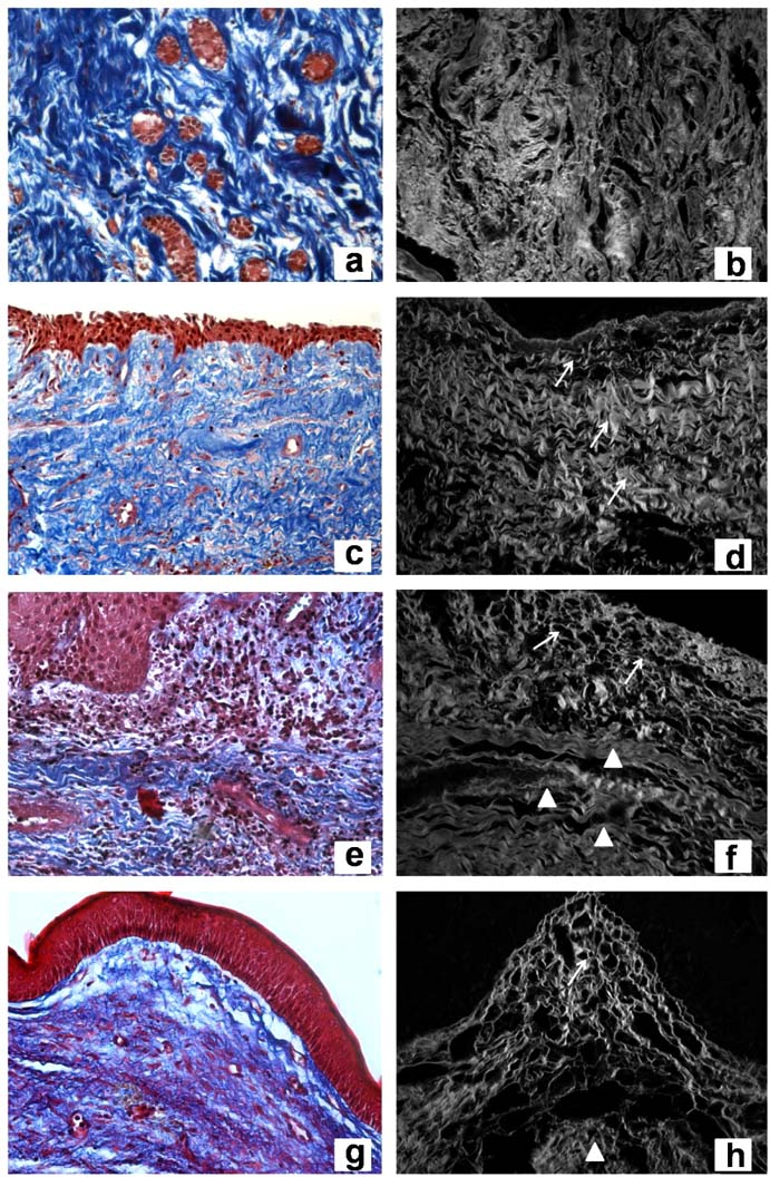
Dentigerous cyst (DC) and keratocystic odontogenic tumor (KOT) are odontogenic lesions arising from epithelial elements, such as those observed in dental follicles (DF), that have been part of the tooth forming apparatus. These lesions show different clinical and histological characteristics, as well as distinct biological behavior. This study aimed to qualify and quantify collagen and elastic fibers by means of histochemical techniques with light and confocal laser microscopic methods in three odontogenic entities. Eleven DF, 13 DC (n=10 with inflammation, n=3 without inflammation) and 13 KOT were processed to the following techniques: Hematoxylin and Eosin, Masson's Trichrome, Picrosirius, Direct Blue, and Orcein. DF and DC without inflammation exhibited collagen with similar characteristics: no parallel pattern of fiber orientation, thick fibers with dense arrangement, and absence of distinct layers. A comparison between DC with inflammation and KOT revealed similar collagen organization, showing distinct layers: thin collagen fibers with loose arrangement near the epithelium and thick fibers with dense arrangement in distant areas. The only difference found was that KOT exhibited a parallel collagen orientation in relation to the odontogenic epithelia. It may be suggested that the connective tissue of DC is a reactive tissue, inducing an expansive growth associated with fluid accumulation and inflammatory process, which in turn may be present as part of the lesion itself. In KOT, loosely arranged collagen may be associated with the behavior of the neoplastic epithelium.
含牙囊肿(DC)和牙源性角化囊性瘤(KOT)是起源于上皮成分的牙源性病变,如在牙囊(DF)中观察到的那些上皮成分,牙囊是牙齿形成器官的一部分。这些病变表现出不同的临床和组织学特征,以及不同的生物学行为。本研究旨在通过组织化学技术结合光镜和共聚焦激光显微镜方法,对三种牙源性病变中的胶原蛋白和弹性纤维进行定性和定量分析。对11个牙囊、13个含牙囊肿(10个有炎症,3个无炎症)和13个牙源性角化囊性瘤进行了以下技术处理:苏木精和伊红染色、Masson三色染色、天狼星红染色、直接蓝染色和地衣红染色。无炎症的牙囊和含牙囊肿表现出相似的胶原蛋白特征:纤维取向无平行模式,纤维粗大且排列密集,无明显分层。有炎症的含牙囊肿与牙源性角化囊性瘤之间的比较显示出相似的胶原蛋白组织,呈现出明显的分层:上皮附近有排列疏松的细胶原纤维,远处有排列密集的粗纤维。唯一的区别是,牙源性角化囊性瘤相对于牙源性上皮表现出平行的胶原取向。可以推测,含牙囊肿的结缔组织是一种反应性组织,诱导与液体聚集和炎症过程相关的膨胀性生长,而炎症过程反过来可能作为病变本身的一部分存在。在牙源性角化囊性瘤中,排列疏松的胶原蛋白可能与肿瘤上皮的行为有关。