Department of Neuroscience and European Graduate School of Neuroscience, Maastricht University, Maastricht, The Netherlands.
Cerebellum. 2012 Mar;11(1):132-44. doi: 10.1007/s12311-011-0297-7.
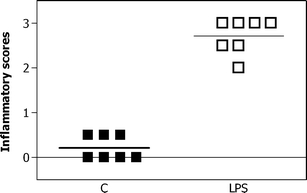
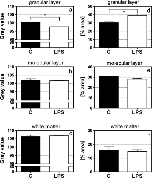
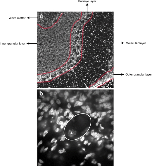
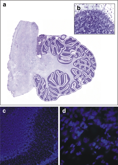
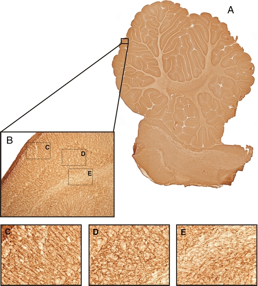
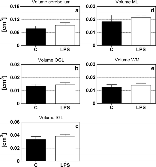
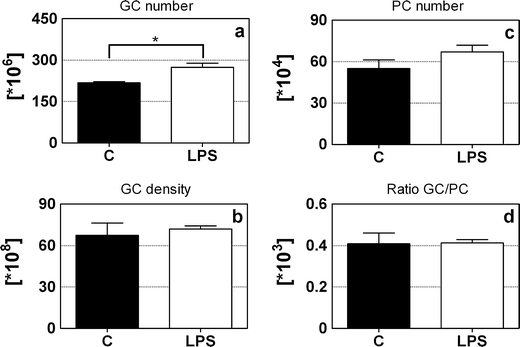
Chorioamnionitis is an important problem in perinatology today, leading to brain injury and neurological handicaps. However, there are almost no data available regarding chorioamnionitis and a specific damage of the cerebellum. Therefore, this study aimed at determining if chorioamnionitis causes cerebellar morphological alterations. Chorioamnionitis was induced in sheep by the intra-amniotic injection of lipopolysaccharide (LPS) at a gestational age (GA) of 110 days. At a GA of 140 days, we assessed the mean total and layer-specific volume and the mean total granule cell (GCs) and Purkinje cell (PC) number in the cerebelli of LPS-exposed and control animals using high-precision design-based stereology. Astrogliosis was assessed in the gray and white matter (WM) using a glial fibrillary acidic protein staining combined with gray value image analysis. The present study showed an unchanged volume of the total cerebellum as well as the molecular layer, outer and inner granular cell layers (OGL and IGL, respectively), and WM. Interestingly, compared with controls, the LPS-exposed brains showed a statistically significant increase (+20.4%) in the mean total number of GCs, whereas the number of PCs did not show any difference between the two groups. In addition, LPS-exposed animals showed signs of astrogliosis specifically affecting the IGL. Intra-amniotic injection of LPS causes morphological changes in the cerebellum of fetal sheep still detectable at full-term birth. In this study, changes were restricted to the inner granule layer. These cerebellar changes might correspond to some of the motor or non-motor deficits seen in neonates from compromised pregnancies.
羊膜腔炎是当今围产医学中的一个重要问题,它可导致脑损伤和神经功能障碍。然而,关于羊膜腔炎和小脑的特定损伤几乎没有数据。因此,本研究旨在确定羊膜腔炎是否会引起小脑形态改变。在胎龄 110 天时,通过向羊膜腔内注射脂多糖(LPS)来诱导羊膜腔炎。在胎龄 140 天时,我们使用高精度基于设计的体视学方法评估 LPS 暴露和对照动物小脑的总平均和分层特异性体积以及总颗粒细胞(GC)和浦肯野细胞(PC)数量。通过胶质纤维酸性蛋白染色结合灰度值图像分析,在灰质和白质(WM)中评估星形胶质细胞增生。本研究显示,总小脑体积以及分子层、外颗粒细胞层(OGL)和内颗粒细胞层(IGL)和 WM 的体积均无变化。有趣的是,与对照组相比,LPS 暴露组的 GC 总数增加了统计学意义(+20.4%),而两组之间 PC 数量没有差异。此外,LPS 暴露的动物表现出星形胶质细胞增生的迹象,特别是影响 IGL。羊膜腔内注射 LPS 会导致胎羊小脑的形态发生变化,这种变化在足月出生时仍可检测到。在本研究中,变化仅限于内颗粒层。这些小脑变化可能与围产期受损的新生儿中出现的一些运动或非运动缺陷相对应。