Mattoli Filippo, Tiribuzi Roberto, D'Angelo Francesco, di Girolamo Ilaria, Quattrocelli Mattia, Montesano Simona, Crispoltoni Lucia, Oikonomou Vasileios, Cusella De Angelis Maria Gabriella, Marconi Peggy, Orlacchio Antonio, Sampaolesi Maurilio, Martino Sabata, Orlacchio Aldo
Dipartimento di Medicina Sperimentale e Scienze Biochimiche, Sezione di Biochimica e Biologia Molecolare, Università degli Studi di Perugia, Via del Giochetto, 06126 Perugia, Italy.
Int J Biomed Imaging. 2011;2011:236854. doi: 10.1155/2011/236854. Epub 2011 Jun 13.
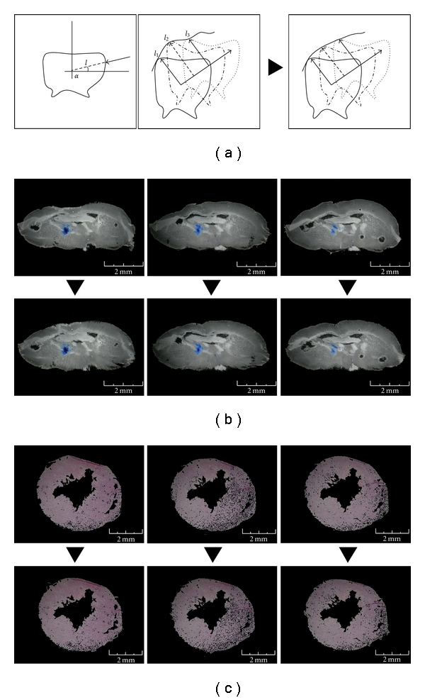
The effectiveness of therapeutic treatment based on regenerative medicine for degenerative diseases (i.e., neurodegenerative or cardiac diseases) requires tools allowing the visualization and analysis of the three-dimensional (3D) distribution of target drugs within the tissue. Here, we present a new computational procedure able to overcome the limitations of visual analysis emerging by the examination of a molecular signal within images of serial tissue/organ sections by using the conventional techniques. Together with the 3D anatomical reconstitution of the tissue/organ, our framework allows the detection of signals of different origins (e.g., marked generic molecules, colorimetric, or fluorimetric substrates for enzymes; microRNA; recombinant protein). Remarkably, the application does not require the employment of specific tracking reagents for the imaging analysis. We report two different representative applications: the first shows the reconstruction of a 3D model of mouse brain with the analysis of the distribution of the β-Galactosidase, the second shows the reconstruction of a 3D mouse heart with the measurement of the cardiac volume.
基于再生医学的退行性疾病(即神经退行性疾病或心脏病)治疗方法的有效性,需要能够对组织内目标药物的三维(3D)分布进行可视化和分析的工具。在此,我们提出一种新的计算程序,该程序能够克服使用传统技术检查连续组织/器官切片图像中的分子信号时出现的视觉分析局限性。连同组织/器官的3D解剖重建,我们的框架允许检测不同来源的信号(例如,标记的通用分子、酶的比色或荧光底物;微小RNA;重组蛋白)。值得注意的是,该应用不需要使用特定的跟踪试剂进行成像分析。我们报告了两种不同的代表性应用:第一个展示了通过分析β-半乳糖苷酶的分布重建小鼠脑的3D模型,第二个展示了通过测量心脏体积重建3D小鼠心脏。