Neuroimaging at MIRA-Institute for Biomedical Technology and Technical Medicine, University of Twente, Enschede, The Netherlands.
PLoS One. 2011;6(7):e22127. doi: 10.1371/journal.pone.0022127. Epub 2011 Jul 13.
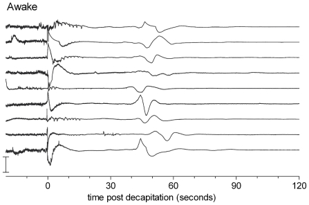
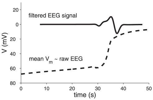
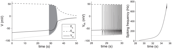
Recent experiments in rats have shown the occurrence of a high amplitude slow brain wave in the EEG approximately 1 minute after decapitation, with a duration of 5-15 s (van Rijn et al, PLoS One 6, e16514, 2011) that was presumed to signify the death of brain neurons. We present a computational model of a single neuron and its intra- and extracellular ion concentrations, which shows the physiological mechanism for this observation. The wave is caused by membrane potential oscillations, that occur after the cessation of activity of the sodium-potassium pumps has lead to an excess of extracellular potassium. These oscillations can be described by the Hodgkin-Huxley equations for the sodium and potassium channels, and result in a sudden change in mean membrane voltage. In combination with a high-pass filter, this sudden depolarization leads to a wave in the EEG. We discuss that this process is not necessarily irreversible.
最近在大鼠身上进行的实验表明,在断头后大约 1 分钟,脑电图中会出现一种高振幅的慢脑波,持续时间为 5-15 秒(van Rijn 等人,PLoS One 6,e16514,2011 年),这被认为是大脑神经元死亡的标志。我们提出了一个单个神经元及其细胞内外离子浓度的计算模型,该模型展示了这一观察结果的生理机制。该波是由细胞膜电位振荡引起的,这种振荡发生在钠钾泵的活动停止导致细胞外钾过多之后。这些振荡可以用钠和钾通道的 Hodgkin-Huxley 方程来描述,并且会导致膜电压的突然变化。与高通滤波器结合使用时,这种去极化的突然发生会导致脑电图中的波。我们讨论了这个过程不一定是不可逆的。