Laboratory of Image Synthesis and Analysis, Faculty of Applied Science, Université Libre de Bruxelles, Brussels, Belgium.
PLoS One. 2011;6(7):e22263. doi: 10.1371/journal.pone.0022263. Epub 2011 Jul 15.
In vitro cell observation has been widely used by biologists and pharmacologists for screening molecule-induced effects on cancer cells. Computer-assisted time-lapse microscopy enables automated live cell imaging in vitro, enabling cell behavior characterization through image analysis, in particular regarding cell migration. In this context, 3D cell assays in transparent matrix gels have been developed to provide more realistic in vitro 3D environments for monitoring cell migration (fundamentally different from cell motility behavior observed in 2D), which is related to the spread of cancer and metastases.
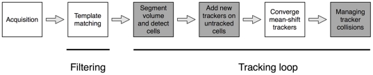
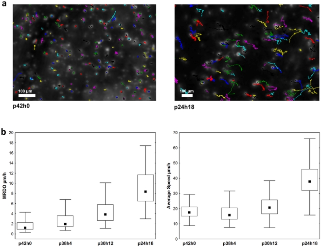
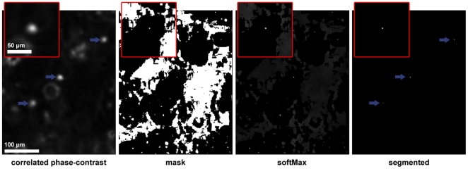
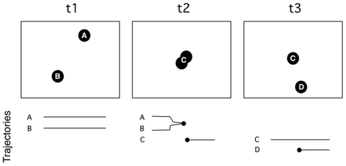
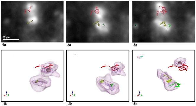
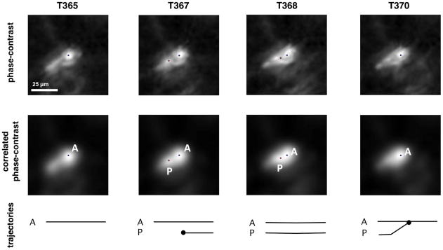
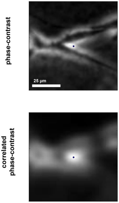
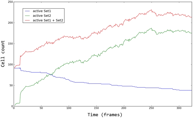
METHODOLOGY/PRINCIPAL FINDINGS: In this paper we propose an improved automated tracking method that is designed to robustly and individually follow a large number of unlabeled cells observed under phase-contrast microscopy in 3D gels. The method automatically detects and tracks individual cells across a sequence of acquired volumes, using a template matching filtering method that in turn allows for robust detection and mean-shift tracking. The robustness of the method results from detecting and managing the cases where two cell (mean-shift) trackers converge to the same point. The resulting trajectories quantify cell migration through statistical analysis of 3D trajectory descriptors. We manually validated the method and observed efficient cell detection and a low tracking error rate (6%). We also applied the method in a real biological experiment where the pro-migratory effects of hyaluronic acid (HA) were analyzed on brain cancer cells. Using collagen gels with increased HA proportions, we were able to evidence a dose-response effect on cell migration abilities.
CONCLUSIONS/SIGNIFICANCE: The developed method enables biomedical researchers to automatically and robustly quantify the pro- or anti-migratory effects of different experimental conditions on unlabeled cell cultures in a 3D environment.
体外细胞观察已被生物学家和药理学家广泛用于筛选分子对癌细胞的诱导作用。计算机辅助时差显微镜使体外自动活细胞成像成为可能,通过图像分析对细胞行为进行特征描述,特别是在细胞迁移方面。在这种情况下,已开发出用于透明基质凝胶的 3D 细胞测定法,以提供更真实的体外 3D 环境来监测细胞迁移(与在 2D 中观察到的细胞迁移行为根本不同),这与癌症和转移的扩散有关。
方法/主要发现:在本文中,我们提出了一种改进的自动跟踪方法,旨在稳健且单独地跟踪在 3D 凝胶中相差显微镜下观察到的大量未标记细胞。该方法使用模板匹配滤波方法自动检测和跟踪各个细胞,该方法又允许稳健检测和均值漂移跟踪。该方法的稳健性源于检测和管理两个细胞(均值漂移)跟踪器收敛到同一位置的情况。通过对 3D 轨迹描述符进行统计分析,得到的轨迹可量化细胞迁移。我们手动验证了该方法,观察到有效的细胞检测和低跟踪错误率(6%)。我们还在真实的生物学实验中应用了该方法,分析了透明质酸(HA)对脑癌细胞的促迁移作用。使用具有增加的 HA 比例的胶原凝胶,我们能够证明细胞迁移能力与剂量反应之间存在关联。
结论/意义:所开发的方法使生物医学研究人员能够自动且稳健地量化不同实验条件对 3D 环境中未标记细胞培养物的促迁移或抗迁移作用。