Ozgoren Murat, Bayazit Onur, Gokmen Necati, Oniz Adile
Department of Biophysics, Faculty of Medicine, Dokuz Eylul University, Izmir Turkey.
Open Neuroimag J. 2010;4:121-9. doi: 10.2174/1874440001004010121. Epub 2010 Sep 8.
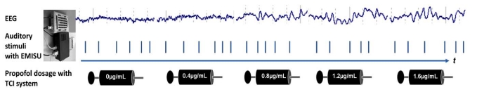
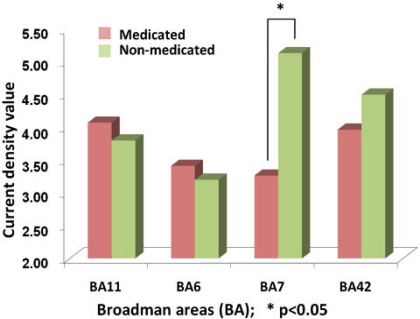
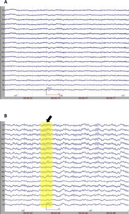
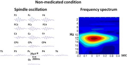
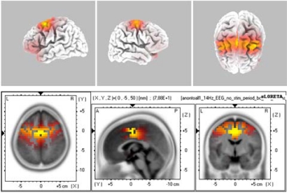
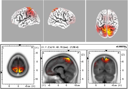
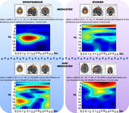
This study's primary objective is to analyze human EEG spindle oscillations during propofol-induced anesthesia and to address possible activation sources. Such an analysis also has a secondary role of investigating the short- term spectral patterns and their functional role.Artifact-free epochs of spindle activations were selected from the electroencephalograms of patients undergoing propofol anesthesia. Power spectral analysis and source localization using standardized low-resolution-brain-electromagnetic-tomography (sLORETA) were performed. Additionally, spectrograms were obtained by means of using the Complex Morlet-based algorithm. In order to highlight the functional properties, auditory stimulations were conducted during the propofol administration. The loss of consciousness was reached at a level of 0.8-1.2 µg/mL, which also provided distinct spindle oscillations in the continuous EEG. The un-evoked (spontaneous) and evoked (auditory) conditions were examined across non-medicated and medicated conditions (propofol). The propofol administration resulted in appearance of 12-14 Hz spindle activity mostly localized in BA6, BA9, BA10, BA21, BA24 and BA37 areas. The presence of auditory stimulations slightly shifted these maximum activities to different locations. Between the medicated and non-medicated conditions, there was a significant reduction of spindle activity, which was pinpointed to BA7 (precuneus area). The findings indicate that spindle oscillations may have a dual nature. That is, spindle oscillations may be activity dependent and disruptive for large-scale information processing networks in the brain. Hence, the study of spindle oscillation may provide a basis for understanding the short-term spectral patterns of human EEG.
本研究的主要目的是分析丙泊酚诱导麻醉期间的人类脑电图纺锤波振荡,并探讨可能的激活源。这种分析还具有调查短期频谱模式及其功能作用的次要作用。从接受丙泊酚麻醉的患者脑电图中选择无伪迹的纺锤波激活时段。进行了功率谱分析和使用标准化低分辨率脑电磁断层扫描(sLORETA)的源定位。此外,通过使用基于复Morlet的算法获得了频谱图。为了突出功能特性,在丙泊酚给药期间进行了听觉刺激。意识丧失发生在0.8 - 1.2 µg/mL的水平,这也在连续脑电图中提供了明显的纺锤波振荡。在未用药和用药(丙泊酚)条件下检查了未诱发(自发)和诱发(听觉)情况。丙泊酚给药导致主要位于BA6、BA9、BA10、BA21、BA24和BA37区域的12 - 14 Hz纺锤波活动出现。听觉刺激的存在使这些最大活动略微转移到不同位置。在用药和未用药条件之间,纺锤波活动显著减少,这一减少集中在BA7(楔前叶区域)。研究结果表明,纺锤波振荡可能具有双重性质。也就是说,纺锤波振荡可能依赖于活动,并对大脑中的大规模信息处理网络具有干扰性。因此,对纺锤波振荡的研究可能为理解人类脑电图的短期频谱模式提供基础。