Department of Chemistry, College of Chemistry and Chemical Engineering, Xiamen University, Xiamen 361005, PR China.
Nanoscale Res Lett. 2011 Jul 29;6(1):480. doi: 10.1186/1556-276X-6-480.
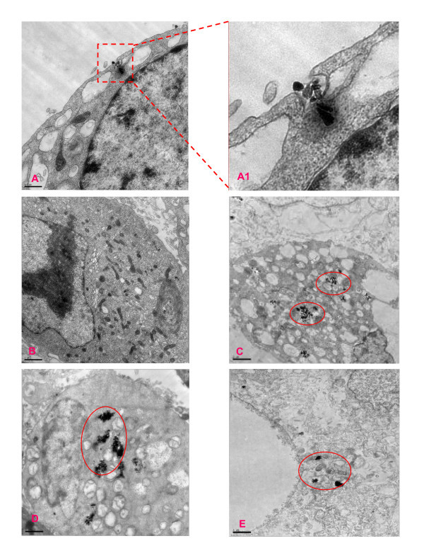
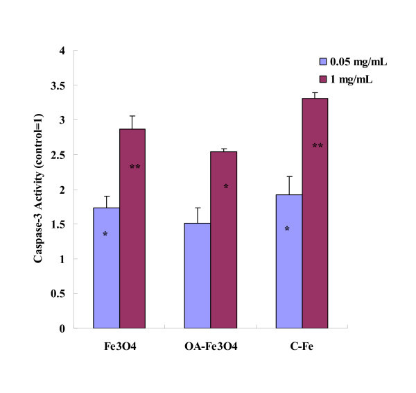
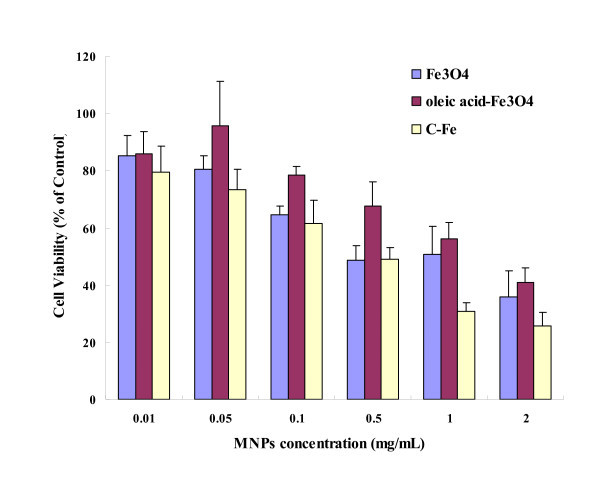
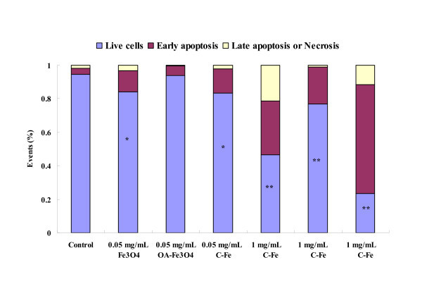
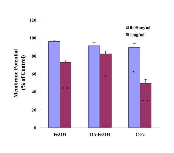
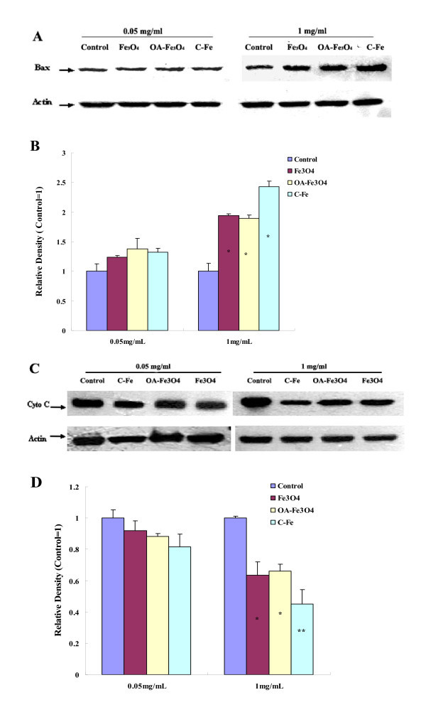
The evaluation of the toxicity of magnetic nanoparticles (MNPs) has attracted much attention in recent years. The current study aimed to investigate the cytotoxic effects of Fe3O4, oleic acid-coated Fe3O4 (OA-Fe3O4), and carbon-coated Fe (C-Fe) nanoparticles on human hepatoma BEL-7402 cells and the mechanisms. WST-1 assay demonstrated that the cytotoxicity of three types of MNPs was in a dose-dependent manner. G1 (Fe3O4 and OA-Fe3O4) phase and G2 (C-Fe) phase cell arrests and apoptosis induced by MNPs were detected by flow cytometry analysis. The increase in apoptosis was accompanied with the Bax over-expression, mitochondrial membrane potential decrease, and the release of cytochrome C from mitochondria into cytosol. Moreover, apoptosis was further confirmed by morphological and biochemical hallmarks, such as swollen mitochondria with lysing cristae and caspase-3 activation. Our results revealed that certain concentrations of the three types of MNPs affect BEL-7402 cells viability via cell arrest and inducing apoptosis, and the MNPs-induced apoptosis is mediated through the mitochondrial-dependent pathway. The influence potency of MNPs observed in all experiments would be: C-Fe > Fe3O4 > OA-Fe3O4.
近年来,磁性纳米粒子(MNPs)的毒性评估引起了广泛关注。本研究旨在探讨不同类型 MNPs(Fe3O4、油酸包覆的 Fe3O4(OA-Fe3O4)和碳包覆的 Fe(C-Fe))对人肝癌 BEL-7402 细胞的细胞毒性及其作用机制。WST-1 法检测结果显示,三种 MNPs 的细胞毒性均呈剂量依赖性。流式细胞术分析结果表明,MNPs 可诱导 BEL-7402 细胞 G1 期(Fe3O4 和 OA-Fe3O4)和 G2 期(C-Fe)阻滞和细胞凋亡。细胞凋亡伴随着 Bax 过表达、线粒体膜电位降低以及细胞色素 C 从线粒体向细胞质的释放。此外,细胞形态学和生化特征进一步证实了细胞凋亡,如线粒体肿胀、嵴溶解和 caspase-3 激活。结果表明,一定浓度的三种 MNPs 通过细胞阻滞和诱导细胞凋亡影响 BEL-7402 细胞活力,且 MNPs 诱导的细胞凋亡是通过线粒体依赖性途径介导的。在所有实验中观察到的 MNPs 影响的效力为:C-Fe > Fe3O4 > OA-Fe3O4。