Department of Psychiatry, University of Magdeburg, Magdeburg, Germany.
J Neuroinflammation. 2011 Aug 10;8:94. doi: 10.1186/1742-2094-8-94.
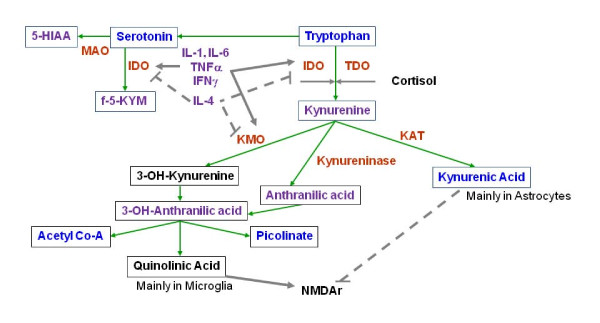
Immune dysfunction, including monocytosis and increased blood levels of interleukin-1, interleukin-6 and tumour necrosis factor α has been observed during acute episodes of major depression. These peripheral immune processes may be accompanied by microglial activation in subregions of the anterior cingulate cortex where depression-associated alterations of glutamatergic neurotransmission have been described.
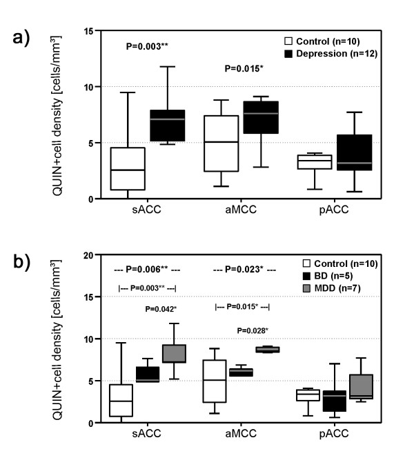
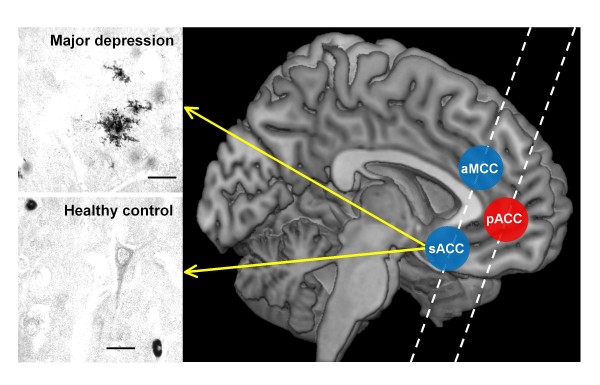
Microglial immunoreactivity of the N-methyl-D-aspartate (NMDA) glutamate receptor agonist quinolinic acid (QUIN) in the subgenual anterior cingulate cortex (sACC), anterior midcingulate cortex (aMCC) and pregenual anterior cingulate cortex (pACC) of 12 acutely depressed suicidal patients (major depressive disorder/MDD, n = 7; bipolar disorder/BD, n = 5) was analyzed using immunohistochemistry and compared with its expression in 10 healthy control subjects.
Depressed patients had a significantly increased density of QUIN-positive cells in the sACC (P = 0.003) and the aMCC (P = 0.015) compared to controls. In contrast, counts of QUIN-positive cells in the pACC did not differ between the groups (P = 0.558). Post-hoc tests showed that significant findings were attributed to MDD and were absent in BD.
These results add a novel link to the immune hypothesis of depression by providing evidence for an upregulation of microglial QUIN in brain regions known to be responsive to infusion of NMDA antagonists such as ketamine. Further work in this area could lead to a greater understanding of the pathophysiology of depressive disorders and pave the way for novel NMDA receptor therapies or immune-modulating strategies.
在重度抑郁症的急性发作期间,观察到免疫功能障碍,包括单核细胞增多症和白细胞介素-1、白细胞介素-6 和肿瘤坏死因子-α 血液水平升高。这些外周免疫过程可能伴随着前扣带回皮质亚区的小胶质细胞激活,在这些区域已经描述了与抑郁相关的谷氨酸能神经传递改变。
使用免疫组织化学分析 12 名急性抑郁自杀患者(重性抑郁障碍/MDD,n=7;双相障碍/BD,n=5)和 10 名健康对照者的 N-甲基-D-天冬氨酸(NMDA)谷氨酸受体激动剂喹啉酸(QUIN)在前扣带皮质亚区(sACC)、前中扣带皮质(aMCC)和前扣带皮质前区(pACC)中的小胶质细胞免疫反应性,并将其与对照组进行比较。
与对照组相比,抑郁患者 sACC(P=0.003)和 aMCC(P=0.015)的 QUIN 阳性细胞密度显著增加。然而,两组间 pACC 的 QUIN 阳性细胞计数无差异(P=0.558)。事后检验显示,显著的发现归因于 MDD,而在 BD 中不存在。
这些结果通过提供证据表明 NMDA 拮抗剂如氯胺酮输注反应性脑区中小胶质细胞 QUIN 的上调,为抑郁症的免疫假说增加了一个新的联系。该领域的进一步研究可能会导致对抑郁障碍病理生理学的更深入理解,并为新型 NMDA 受体治疗或免疫调节策略铺平道路。