• 文献检索
  • 文档翻译
  • 深度研究
  • 学术资讯
  • Suppr Zotero 插件Zotero 插件
  • 邀请有礼
  • 套餐&价格
  • 历史记录
应用&插件
Suppr Zotero 插件Zotero 插件浏览器插件Mac 客户端Windows 客户端微信小程序
定价
高级版会员购买积分包购买API积分包
服务
文献检索文档翻译深度研究API 文档MCP 服务
关于我们
关于 Suppr公司介绍联系我们用户协议隐私条款
关注我们

Suppr 超能文献

核心技术专利:CN118964589B侵权必究
粤ICP备2023148730 号-1Suppr @ 2026

文献检索

告别复杂PubMed语法,用中文像聊天一样搜索,搜遍4000万医学文献。AI智能推荐,让科研检索更轻松。

立即免费搜索

文件翻译

保留排版,准确专业,支持PDF/Word/PPT等文件格式,支持 12+语言互译。

免费翻译文档

深度研究

AI帮你快速写综述,25分钟生成高质量综述,智能提取关键信息,辅助科研写作。

立即免费体验

乳腺 X 光片中的胸肌识别。

Pectoral muscle identification in mammograms.

机构信息

Department of Computer Science and Engineering, National Institute of Technology Calicut, Calicut, India.

出版信息

J Appl Clin Med Phys. 2011 Mar 3;12(3):3285. doi: 10.1120/jacmp.v12i3.3285.

DOI:10.1120/jacmp.v12i3.3285
PMID:21844845
原文链接:https://pmc.ncbi.nlm.nih.gov/articles/PMC5718641/
Abstract

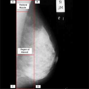
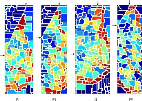
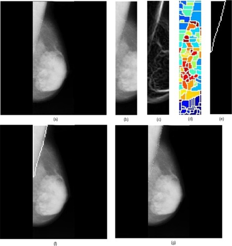
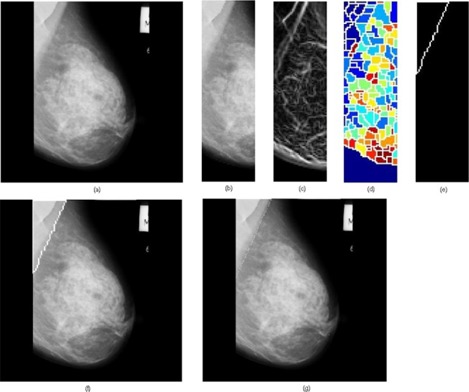
In most of the approaches of computer-aided detection of breast cancer, one of the preprocessing steps applied to the mammogram is the removal/suppression of pectoral muscle, as its presence within the mammogram may adversely affect the outcome of cancer detection processes. Through this study, we propose an efficient automatic method using the watershed transformation for identifying the pectoral muscle in mediolateral oblique view mammograms. The watershed transformation of the mammogram shows interesting properties that include the appearance of a unique watershed line corresponding to the pectoral muscle edge. In addition to this, it is observed that the pectoral muscle region is oversegmented due to the existence of several catchment basins within the pectoral muscle. Hence, a suitable merging algorithm is proposed to combine the appropriate catchment basins to obtain the correct pectoral muscle region. A total of 84 mammograms from the mammographic image analysis database were used to validate this approach. The mean false positive and mean false negative rates, obtained by comparing the results of the proposed approach with manually-identified (ground truth) pectoral muscle boundaries, respectively, were 0.85% and 4.88%. A comparison of the results of the proposed method with related state-of-the-art methods shows that the performance of the proposed approach is better than the existing methods in terms of the mean false negative rate. Using Hausdorff distance metric, the comparison of the results of the proposed method with ground truth shows low Hausdorff distances, the mean and standard deviation being 3.85 ± 1.07 mm.

摘要

在大多数计算机辅助乳腺癌检测方法中,应用于乳房 X 光片的预处理步骤之一是去除/抑制胸肌,因为其存在于乳房 X 光片中可能会对癌症检测过程的结果产生不利影响。通过这项研究,我们提出了一种使用分水岭变换识别中侧斜位乳房 X 光片中胸肌的有效自动方法。乳房 X 光片的分水岭变换具有有趣的特性,包括出现与胸肌边缘对应的独特分水岭线。除此之外,还观察到由于胸肌内存在多个集水盆地,胸肌区域被过度分割。因此,提出了一种合适的合并算法来合并适当的集水盆地,以获得正确的胸肌区域。该方法共使用了来自乳腺图像分析数据库的 84 张乳房 X 光片进行验证。通过将所提出方法的结果与手动识别(地面实况)的胸肌边界进行比较,分别获得的平均假阳性和平均假阴性率分别为 0.85%和 4.88%。将所提出方法的结果与相关的最先进方法进行比较表明,在所提出方法的平均假阴性率方面,其性能优于现有方法。使用 Hausdorff 距离度量,将所提出方法的结果与地面实况进行比较显示出较低的 Hausdorff 距离,平均值和标准差分别为 3.85±1.07mm。

https://cdn.ncbi.nlm.nih.gov/pmc/blobs/0093/5718641/b7d6a42e0a55/ACM2-12-215-g008.jpg
https://cdn.ncbi.nlm.nih.gov/pmc/blobs/0093/5718641/8a4578c8fec5/ACM2-12-215-g002.jpg
https://cdn.ncbi.nlm.nih.gov/pmc/blobs/0093/5718641/1814cf99a71e/ACM2-12-215-g004.jpg
https://cdn.ncbi.nlm.nih.gov/pmc/blobs/0093/5718641/2c6e12ebadb1/ACM2-12-215-g006.jpg
https://cdn.ncbi.nlm.nih.gov/pmc/blobs/0093/5718641/c1b45364362e/ACM2-12-215-g007.jpg
https://cdn.ncbi.nlm.nih.gov/pmc/blobs/0093/5718641/b7d6a42e0a55/ACM2-12-215-g008.jpg

相似文献

1
Pectoral muscle identification in mammograms.乳腺 X 光片中的胸肌识别。
J Appl Clin Med Phys. 2011 Mar 3;12(3):3285. doi: 10.1120/jacmp.v12i3.3285.
2
Computer-aided identification of the pectoral muscle in digitized mammograms.数字化乳腺 X 线片中胸大肌的计算机辅助识别。
J Digit Imaging. 2010 Oct;23(5):562-80. doi: 10.1007/s10278-009-9240-6. Epub 2009 Oct 9.
3
Detection and Segmentation of Pectoral Muscle on MLO-View Mammogram Using Enhancement Filter.基于增强滤波器的 MLO 视图乳腺钼靶图像中胸肌的检测与分割。
J Med Syst. 2017 Oct 25;41(12):190. doi: 10.1007/s10916-017-0839-8.
4
Automatic identification of the pectoral muscle in mammograms.乳腺钼靶片中胸肌的自动识别。
IEEE Trans Med Imaging. 2004 Feb;23(2):232-45. doi: 10.1109/tmi.2003.823062.
5
Robust Automatic Pectoral Muscle Segmentation from Mammograms Using Texture Gradient and Euclidean Distance Regression.使用纹理梯度和欧几里得距离回归从乳房X光片中进行稳健的自动胸肌分割
J Digit Imaging. 2016 Feb;29(1):115-25. doi: 10.1007/s10278-015-9813-5.
6
Geometry-Based Pectoral Muscle Segmentation From MLO Mammogram Views.基于几何方法的MLO位乳房X线照片视图中胸肌分割
IEEE Trans Biomed Eng. 2017 Nov;64(11):2662-2671. doi: 10.1109/TBME.2017.2649481.
7
A robust method for segmenting pectoral muscle in mediolateral oblique (MLO) mammograms.一种用于在斜侧位(MLO)乳房 X 光片中分割胸肌的稳健方法。
Int J Comput Assist Radiol Surg. 2019 Feb;14(2):237-248. doi: 10.1007/s11548-018-1867-7. Epub 2018 Oct 4.
8
Automatic Detection of Pectoral Muscle Region for Computer-Aided Diagnosis Using MIAS Mammograms.使用MIAS乳腺X线照片进行计算机辅助诊断时胸肌区域的自动检测
Biomed Res Int. 2016;2016:5967580. doi: 10.1155/2016/5967580. Epub 2016 Oct 25.
9
Automated pectoral muscle identification on MLO-view mammograms: Comparison of deep neural network to conventional computer vision.基于 MLO 视图的乳腺钼靶片中自动胸大肌识别:深度神经网络与传统计算机视觉的比较。
Med Phys. 2019 May;46(5):2103-2114. doi: 10.1002/mp.13451. Epub 2019 Mar 12.
10
Automatic pectoral muscle segmentation on mediolateral oblique view mammograms.在内外侧斜位乳腺钼靶片上自动分割胸肌
IEEE Trans Med Imaging. 2004 Sep;23(9):1129-40. doi: 10.1109/TMI.2004.830529.

引用本文的文献

1
Automatic Pectoral Muscle Region Segmentation in Mammograms Using Genetic Algorithm and Morphological Selection.基于遗传算法和形态学选择的乳腺 X 光片中胸肌区域自动分割。
J Digit Imaging. 2018 Oct;31(5):680-691. doi: 10.1007/s10278-018-0068-9.
2
Automatic Detection of Pectoral Muscle Region for Computer-Aided Diagnosis Using MIAS Mammograms.使用MIAS乳腺X线照片进行计算机辅助诊断时胸肌区域的自动检测
Biomed Res Int. 2016;2016:5967580. doi: 10.1155/2016/5967580. Epub 2016 Oct 25.
3
Identification and segmentation of obscure pectoral muscle in mediolateral oblique mammograms.

本文引用的文献

1
Computer-aided identification of the pectoral muscle in digitized mammograms.数字化乳腺 X 线片中胸大肌的计算机辅助识别。
J Digit Imaging. 2010 Oct;23(5):562-80. doi: 10.1007/s10278-009-9240-6. Epub 2009 Oct 9.
2
Computer-aided detection and diagnosis of breast cancer with mammography: recent advances.乳腺钼靶摄影术对乳腺癌的计算机辅助检测与诊断:最新进展
IEEE Trans Inf Technol Biomed. 2009 Mar;13(2):236-51. doi: 10.1109/TITB.2008.2009441. Epub 2009 Jan 20.
3
Automatic pectoral muscle segmentation on mediolateral oblique view mammograms.
中外侧斜位乳腺钼靶片中隐匿胸肌的识别与分割
Br J Radiol. 2016 Jun;89(1062):20150802. doi: 10.1259/bjr.20150802. Epub 2016 Apr 4.
4
Review of recent advances in segmentation of the breast boundary and the pectoral muscle in mammograms.乳腺钼靶片中乳房边界和胸肌分割的最新进展综述。
Med Biol Eng Comput. 2016 Jul;54(7):1003-24. doi: 10.1007/s11517-015-1411-7. Epub 2015 Nov 6.
5
Shape-based Automatic Detection of Pectoral Muscle Boundary in Mammograms.基于形状的乳房X光片中胸肌边界自动检测
J Med Biol Eng. 2015;35(3):315-322. doi: 10.1007/s40846-015-0043-6. Epub 2015 Jun 10.
在内外侧斜位乳腺钼靶片上自动分割胸肌
IEEE Trans Med Imaging. 2004 Sep;23(9):1129-40. doi: 10.1109/TMI.2004.830529.
4
Improved watershed transform for medical image segmentation using prior information.利用先验信息改进分水岭变换用于医学图像分割
IEEE Trans Med Imaging. 2004 Apr;23(4):447-58. doi: 10.1109/TMI.2004.824224.
5
Automatic identification of the pectoral muscle in mammograms.乳腺钼靶片中胸肌的自动识别。
IEEE Trans Med Imaging. 2004 Feb;23(2):232-45. doi: 10.1109/tmi.2003.823062.
6
Three-dimensional reconstruction of microcalcification clusters from two mammographic views.从两个乳腺钼靶视图对微钙化簇进行三维重建。
IEEE Trans Med Imaging. 2001 Jun;20(6):479-89.
7
Automated classification of parenchymal patterns in mammograms.乳腺钼靶片中实质模式的自动分类。
Phys Med Biol. 1998 Feb;43(2):365-78. doi: 10.1088/0031-9155/43/2/011.
8
Segmentation of mammograms using multiple linked self-organizing neural networks.使用多个链接自组织神经网络对乳房X光照片进行分割
Med Phys. 1995 Feb;22(2):145-52. doi: 10.1118/1.597464.