INRA, UMR444 Génétique Cellulaire, Auzeville, Castanet-Tolosan, France.
BMC Genomics. 2011 Aug 18;12:417. doi: 10.1186/1471-2164-12-417.
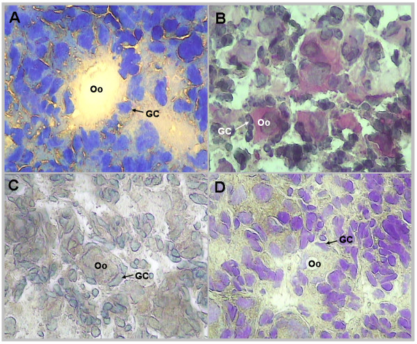
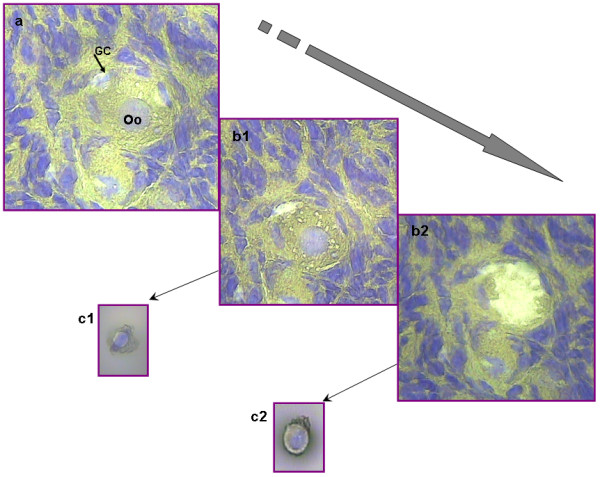
Successful achievement of early folliculogenesis is crucial for female reproductive function. The process is finely regulated by cell-cell interactions and by the coordinated expression of genes in both the oocyte and in granulosa cells. Despite many studies, little is known about the cell-specific gene expression driving early folliculogenesis. The very small size of these follicles and the mixture of types of follicles within the developing ovary make the experimental study of isolated follicular components very difficult.The recently developed laser capture microdissection (LCM) technique coupled with microarray experiments is a promising way to address the molecular profile of pure cell populations. However, one main challenge was to preserve the RNA quality during the isolation of single cells or groups of cells and also to obtain sufficient amounts of RNA.Using a new LCM method, we describe here the separate expression profiles of oocytes and follicular cells during the first stages of sheep folliculogenesis.
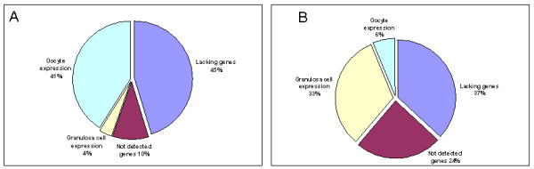
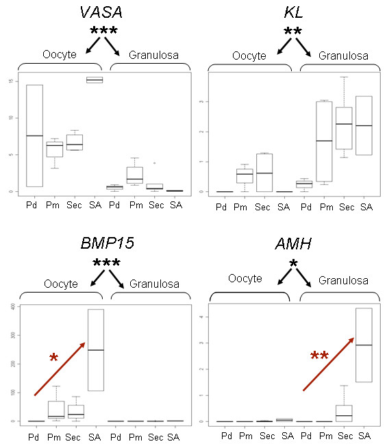
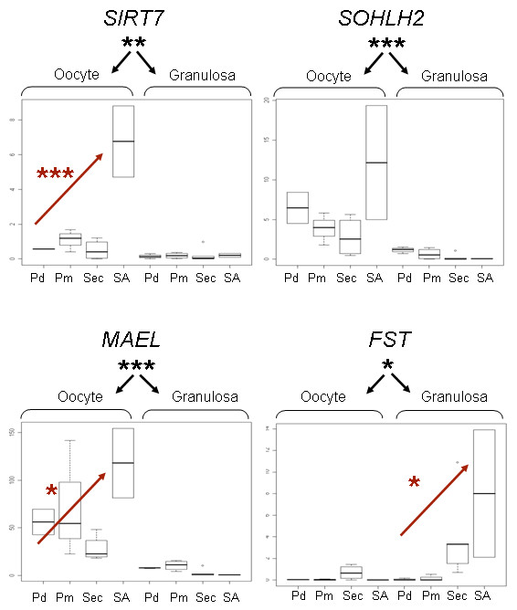
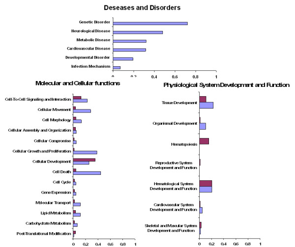
We developed a new tissue fixation protocol ensuring efficient single cell capture and RNA integrity during the microdissection procedure. Enrichment in specific cell types was controlled by qRT-PCR analysis of known genes: six oocyte-specific genes (SOHLH2, MAEL, MATER, VASA, GDF9, BMP15) and three granulosa cell-specific genes (KL, GATA4, AMH).A global gene expression profile for each follicular compartment during early developmental stages was identified here for the first time, using a bovine Affymetrix chip. Most notably, the granulosa cell dataset is unique to date. The comparison of oocyte vs. follicular cell transcriptomes revealed 1050 transcripts specific to the granulosa cell and 759 specific to the oocyte.Functional analyses allowed the characterization of the three main cellular events involved in early folliculogenesis and confirmed the relevance and potential of LCM-derived RNA.
The ovary is a complex mixture of different cell types. Distinct cell populations need therefore to be analyzed for a better understanding of their potential interactions. LCM and microarray analysis allowed us to identify novel gene expression patterns in follicular cells at different stages and in oocyte populations.
早期卵泡发生的成功实现对女性生殖功能至关重要。该过程受到细胞-细胞相互作用以及卵母细胞和颗粒细胞中基因协调表达的精细调节。尽管进行了许多研究,但对于驱动早期卵泡发生的细胞特异性基因表达知之甚少。这些卵泡非常小,并且在发育中的卵巢中混合了各种类型的卵泡,这使得对分离的卵泡成分进行实验研究变得非常困难。最近开发的激光捕获显微解剖 (LCM) 技术与微阵列实验相结合,是解决纯细胞群体分子特征的一种很有前途的方法。然而,一个主要挑战是在分离单个细胞或细胞群时保持 RNA 质量,并且还要获得足够量的 RNA。在这里,我们使用一种新的 LCM 方法描述了绵羊卵泡发生早期阶段卵母细胞和卵泡细胞的单独表达谱。
我们开发了一种新的组织固定方案,可确保在微解剖过程中有效捕获单个细胞并保持 RNA 完整性。通过对已知基因(SOHLH2、MAEL、MATER、VASA、GDF9、BMP15)的 qRT-PCR 分析,以及三个颗粒细胞特异性基因(KL、GATA4、AMH),控制特定细胞类型的富集。在这里,我们首次使用牛 Affymetrix 芯片为每个卵泡隔室确定了早期发育阶段的全局基因表达谱。值得注意的是,颗粒细胞数据集是迄今为止独一无二的。卵母细胞与卵泡细胞转录组的比较揭示了 1050 个转录本是颗粒细胞特异性的,759 个转录本是卵母细胞特异性的。功能分析允许对涉及早期卵泡发生的三个主要细胞事件进行特征描述,并证实了 LCM 衍生 RNA 的相关性和潜力。
卵巢是不同细胞类型的复杂混合物。因此,需要分析不同的细胞群体,以更好地了解它们的潜在相互作用。LCM 和微阵列分析使我们能够识别不同阶段卵泡细胞和卵母细胞群体中的新基因表达模式。