• 文献检索
  • 文档翻译
  • 深度研究
  • 学术资讯
  • Suppr Zotero 插件Zotero 插件
  • 邀请有礼
  • 套餐&价格
  • 历史记录
应用&插件
Suppr Zotero 插件Zotero 插件浏览器插件Mac 客户端Windows 客户端微信小程序
定价
高级版会员购买积分包购买API积分包
服务
文献检索文档翻译深度研究API 文档MCP 服务
关于我们
关于 Suppr公司介绍联系我们用户协议隐私条款
关注我们

Suppr 超能文献

核心技术专利:CN118964589B侵权必究
粤ICP备2023148730 号-1Suppr @ 2026

文献检索

告别复杂PubMed语法,用中文像聊天一样搜索,搜遍4000万医学文献。AI智能推荐,让科研检索更轻松。

立即免费搜索

文件翻译

保留排版,准确专业,支持PDF/Word/PPT等文件格式,支持 12+语言互译。

免费翻译文档

深度研究

AI帮你快速写综述,25分钟生成高质量综述,智能提取关键信息,辅助科研写作。

立即免费体验

dlx 和 sp6-9 控制原型眼的视杯再生。

dlx and sp6-9 Control optic cup regeneration in a prototypic eye.

机构信息

Whitehead Institute for Biomedical Research, Cambridge, Massachusetts, United States of America.

出版信息

PLoS Genet. 2011 Aug;7(8):e1002226. doi: 10.1371/journal.pgen.1002226. Epub 2011 Aug 11.

DOI:10.1371/journal.pgen.1002226
PMID:21852957
原文链接:https://pmc.ncbi.nlm.nih.gov/articles/PMC3154955/
Abstract

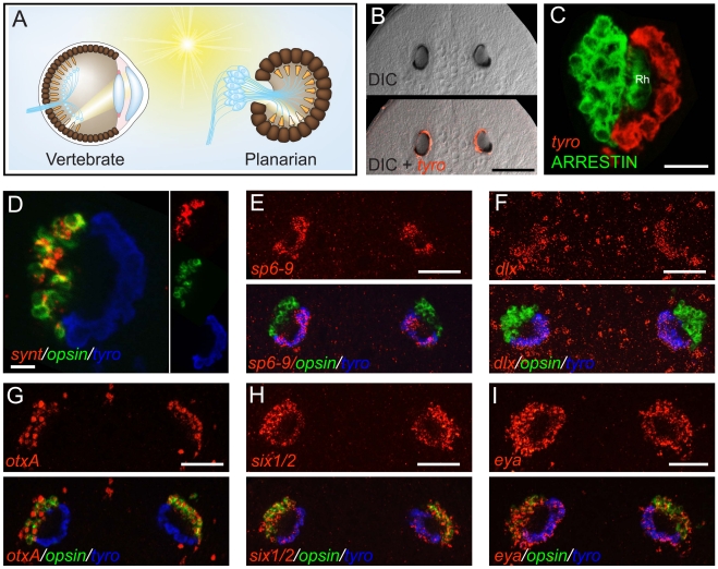
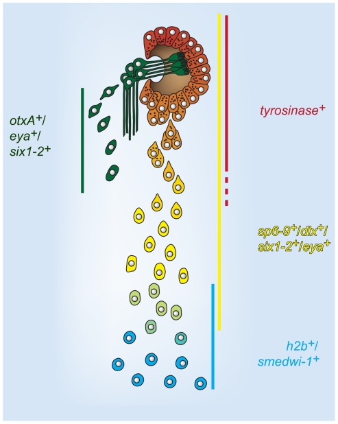
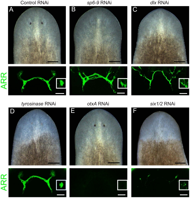
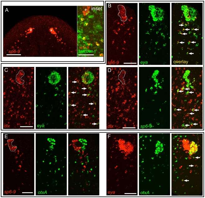
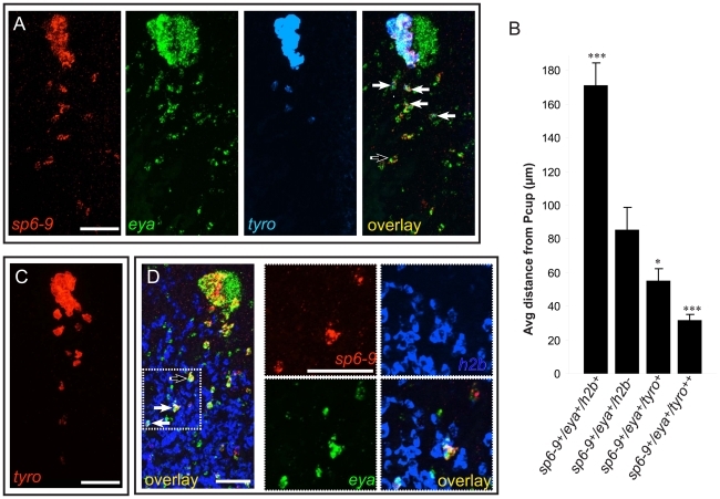
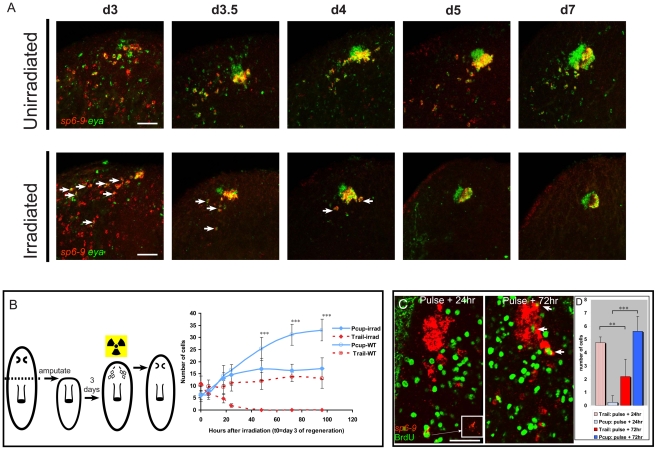
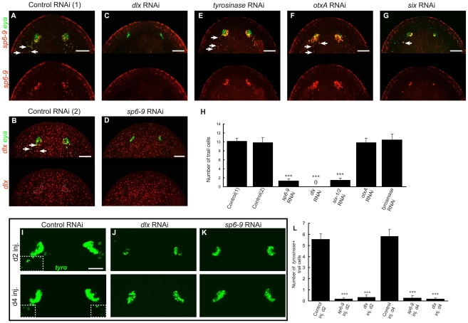
Optic cups are a structural feature of diverse eyes, from simple pit eyes to camera eyes of vertebrates and cephalopods. We used the planarian prototypic eye as a model to study the genetic control of optic cup formation and regeneration. We identified two genes encoding transcription factors, sp6-9 and dlx, that were expressed in the eye specifically in the optic cup and not the photoreceptor neurons. RNAi of these genes prevented formation of visible optic cups during regeneration. Planarian regeneration requires an adult proliferative cell population with stem cell-like properties called the neoblasts. We found that optic cup formation occurred only after migration of progressively differentiating progenitor cells from the neoblast population. The eye regeneration defect caused by dlx and sp6-9 RNAi can be explained by a failure to generate these early optic cup progenitors. Dlx and Sp6-9 genes function as a module during the development of diverse animal appendages, including vertebrate and insect limbs. Our work reveals a novel function for this gene pair in the development of a fundamental eye component, and it utilizes these genes to demonstrate a mechanism for total organ regeneration in which extensive cell movement separates new cell specification from organ morphogenesis.

摘要

视杯是多种眼睛的结构特征,从简单的眼窝到脊椎动物和头足类动物的摄像头眼。我们使用扁形虫原型眼作为模型,研究视杯形成和再生的遗传控制。我们鉴定了两个编码转录因子 sp6-9 和 dlx 的基因,这些基因在眼睛中特异性地在视杯中表达,而不在光感受器神经元中表达。这些基因的 RNAi 阻止了再生过程中可见视杯的形成。扁形虫再生需要具有成体增殖细胞特性的细胞群体,称为成体干细胞样细胞,即 neoblasts。我们发现,只有当来自 neoblast 群体的逐渐分化的祖细胞迁移时,视杯的形成才会发生。dlx 和 sp6-9 RNAi 引起的眼睛再生缺陷可以通过未能产生这些早期视杯祖细胞来解释。Dlx 和 Sp6-9 基因在包括脊椎动物和昆虫肢体在内的各种动物附肢的发育中作为一个模块发挥作用。我们的工作揭示了这对基因在基本眼睛成分发育中的新功能,并利用这些基因证明了一种用于总器官再生的机制,其中广泛的细胞运动将新的细胞特化与器官形态发生分开。

https://cdn.ncbi.nlm.nih.gov/pmc/blobs/0c0b/3154955/9c6b3aa52771/pgen.1002226.g007.jpg
https://cdn.ncbi.nlm.nih.gov/pmc/blobs/0c0b/3154955/2a85fa528b62/pgen.1002226.g001.jpg
https://cdn.ncbi.nlm.nih.gov/pmc/blobs/0c0b/3154955/5b79b9fdbe6d/pgen.1002226.g002.jpg
https://cdn.ncbi.nlm.nih.gov/pmc/blobs/0c0b/3154955/be202624a673/pgen.1002226.g003.jpg
https://cdn.ncbi.nlm.nih.gov/pmc/blobs/0c0b/3154955/85ae8d1f2c12/pgen.1002226.g004.jpg
https://cdn.ncbi.nlm.nih.gov/pmc/blobs/0c0b/3154955/4b525871a239/pgen.1002226.g005.jpg
https://cdn.ncbi.nlm.nih.gov/pmc/blobs/0c0b/3154955/68b9027e6a71/pgen.1002226.g006.jpg
https://cdn.ncbi.nlm.nih.gov/pmc/blobs/0c0b/3154955/9c6b3aa52771/pgen.1002226.g007.jpg

相似文献

1
dlx and sp6-9 Control optic cup regeneration in a prototypic eye.dlx 和 sp6-9 控制原型眼的视杯再生。
PLoS Genet. 2011 Aug;7(8):e1002226. doi: 10.1371/journal.pgen.1002226. Epub 2011 Aug 11.
2
Smed-egfr-4 is required for planarian eye regeneration.涡虫眼再生需要Smed-egfr-4。
Int J Dev Biol. 2019;63(1-2):9-15. doi: 10.1387/ijdb.180361fc.
3
Transcriptome analysis of the planarian eye identifies ovo as a specific regulator of eye regeneration.涡虫眼转录组分析鉴定ovo 为眼再生的特定调控因子。
Cell Rep. 2012 Aug 30;2(2):294-307. doi: 10.1016/j.celrep.2012.06.018. Epub 2012 Aug 2.
4
Specialized progenitors and regeneration.特化祖细胞与再生。
Development. 2013 Mar;140(5):951-7. doi: 10.1242/dev.080499.
5
[Molecular approach to planarian stem cell system].[涡虫干细胞系统的分子研究方法]
Tanpakushitsu Kakusan Koso. 2005 May;50(6 Suppl):706-10.
6
Self-organization and progenitor targeting generate stable patterns in planarian regeneration.自组织和祖细胞靶向在扁形动物再生中产生稳定的模式。
Science. 2018 Apr 27;360(6387):404-409. doi: 10.1126/science.aap8179. Epub 2018 Mar 15.
7
SMEDWI-2 is a PIWI-like protein that regulates planarian stem cells.SMEDWI-2是一种调控涡虫干细胞的类PIWI蛋白。
Science. 2005 Nov 25;310(5752):1327-30. doi: 10.1126/science.1116110.
8
Cellular and molecular dissection of pluripotent adult somatic stem cells in planarians.涡虫多能成体干细胞的细胞和分子剖析。
Dev Growth Differ. 2010 Jan;52(1):27-41. doi: 10.1111/j.1440-169X.2009.01155.x.
9
Neoblast-enriched zinc finger protein FIR1 triggers local proliferation during planarian regeneration.富含成体干细胞的锌指蛋白 FIR1 触发涡虫再生过程中的局部增殖。
Protein Cell. 2019 Jan;10(1):43-59. doi: 10.1007/s13238-018-0512-0. Epub 2018 Mar 20.
10
A regulatory program for excretory system regeneration in planarians.扁形动物排泄系统再生的调控程序。
Development. 2011 Oct;138(20):4387-98. doi: 10.1242/dev.068098.

引用本文的文献

1
A parenchymal niche regulates pluripotent stem cell function in planarians.实质小生境调节涡虫中多能干细胞的功能。
bioRxiv. 2025 Aug 2:2025.08.01.668211. doi: 10.1101/2025.08.01.668211.
2
Developmental onset of planarian whole-body regeneration depends on axis reset.涡虫全身再生的发育起始取决于轴重设。
Curr Biol. 2025 Jun 9;35(11):2479-2494.e3. doi: 10.1016/j.cub.2025.03.065. Epub 2025 Apr 15.
3
map3k1 suppresses terminal differentiation of migratory eye progenitors in planarian regeneration.Map3k1抑制涡虫再生过程中迁移性眼祖细胞的终末分化。

本文引用的文献

1
Clonogenic neoblasts are pluripotent adult stem cells that underlie planarian regeneration.克隆性成体神经细胞是多能性成年干细胞,是扁形动物再生的基础。
Science. 2011 May 13;332(6031):811-6. doi: 10.1126/science.1203983.
2
Planarian regeneration involves distinct stem cell responses to wounds and tissue absence.扁形动物再生涉及到对创伤和组织缺失的不同干细胞反应。
Dev Biol. 2010 Aug 15;344(2):979-91. doi: 10.1016/j.ydbio.2010.06.017. Epub 2010 Jun 19.
3
Non-redundant selector and growth-promoting functions of two sister genes, buttonhead and Sp1, in Drosophila leg development.
PLoS Genet. 2025 Mar 17;21(3):e1011457. doi: 10.1371/journal.pgen.1011457. eCollection 2025 Mar.
4
Stem cells (neoblasts) and positional information jointly dominate regeneration in planarians.干细胞(新生细胞)和位置信息共同主导涡虫的再生过程。
Heliyon. 2025 Jan 9;11(2):e41833. doi: 10.1016/j.heliyon.2025.e41833. eCollection 2025 Jan 30.
5
Reduced adult stem cell fate specification led to eye reduction in cave planarians.成年干细胞命运特化的减少导致洞穴涡虫眼睛缩小。
Nat Commun. 2025 Jan 2;16(1):304. doi: 10.1038/s41467-024-54478-6.
6
suppresses terminal differentiation of migratory eye progenitors in planarian regeneration.抑制涡虫再生过程中迁移性眼祖细胞的终末分化。
bioRxiv. 2024 Oct 12:2024.10.11.617745. doi: 10.1101/2024.10.11.617745.
7
SoxC and MmpReg promote blastema formation in whole-body regeneration of fragmenting potworms Enchytraeus japonensis.SoxC 和 MmpReg 促进碎裂涡虫整体再生中的芽基形成。
Nat Commun. 2024 Aug 22;15(1):6659. doi: 10.1038/s41467-024-50865-1.
8
Mechanistic regulation of planarian shape during growth and degrowth.在生长和萎缩过程中扁形动物形状的机制调节。
Development. 2024 May 1;151(9). doi: 10.1242/dev.202353. Epub 2024 May 9.
9
A transcription factor atlas of stem cell fate in planarians.转录因子图谱:扁形动物干细胞命运决定。
Cell Rep. 2024 Mar 26;43(3):113843. doi: 10.1016/j.celrep.2024.113843. Epub 2024 Feb 23.
10
LIM-HD transcription factors control axial patterning and specify distinct neuronal and intestinal cell identities in planarians.LIM-HD 转录因子控制扁形动物的轴向模式,并在扁形动物中特异性指定不同的神经元和肠道细胞身份。
Open Biol. 2023 Dec;13(12):230327. doi: 10.1098/rsob.230327. Epub 2023 Dec 13.
在果蝇腿发育过程中,两个姐妹基因 buttonhead 和 Sp1 具有非冗余的选择器和促进生长的功能。
PLoS Genet. 2010 Jun 24;6(6):e1001001. doi: 10.1371/journal.pgen.1001001.
4
Co-option of an anteroposterior head axis patterning system for proximodistal patterning of appendages in early bilaterian evolution.头前后轴模式系统在早期两侧对称动物进化中对附肢近-远轴模式的兼用。
Dev Biol. 2010 Aug 1;344(1):358-62. doi: 10.1016/j.ydbio.2010.04.022. Epub 2010 May 8.
5
A clustered set of three Sp-family genes is ancestral in the Metazoa: evidence from sequence analysis, protein domain structure, developmental expression patterns and chromosomal location.在后生动物中,Sp 家族的三个基因簇是祖先的:来自序列分析、蛋白质结构域、发育表达模式和染色体定位的证据。
BMC Evol Biol. 2010 Mar 30;10:88. doi: 10.1186/1471-2148-10-88.
6
Single-cell gene profiling of planarian stem cells using fluorescent activated cell sorting and its "index sorting" function for stem cell research.利用荧光激活细胞分选对涡虫干细胞进行单细胞基因谱分析及其在干细胞研究中的“索引分选”功能。
Dev Growth Differ. 2010 Jan;52(1):131-44. doi: 10.1111/j.1440-169X.2009.01157.x.
7
Planarians maintain a constant ratio of different cell types during changes in body size by using the stem cell system.涡虫通过利用干细胞系统,在身体大小发生变化时维持不同细胞类型的恒定比例。
Zoolog Sci. 2009 Dec;26(12):805-13. doi: 10.2108/zsj.26.805.
8
Role of Epiprofin, a zinc-finger transcription factor, in limb development.Epiprofin,一种锌指转录因子,在肢体发育中的作用。
Dev Biol. 2010 Jan 15;337(2):363-74. doi: 10.1016/j.ydbio.2009.11.007. Epub 2009 Nov 10.
9
A wound-induced Wnt expression program controls planarian regeneration polarity.伤口诱导的 Wnt 表达程序控制扁形动物再生的极性。
Proc Natl Acad Sci U S A. 2009 Oct 6;106(40):17061-6. doi: 10.1073/pnas.0906823106. Epub 2009 Sep 14.
10
A conserved function of the zinc finger transcription factor Sp8/9 in allometric appendage growth in the milkweed bug Oncopeltus fasciatus.锌指转录因子 Sp8/9 在马利筋盲蝽所有附肢生长中的保守功能。
Dev Genes Evol. 2009 Aug;219(8):427-35. doi: 10.1007/s00427-009-0301-0. Epub 2009 Sep 16.