• 文献检索
  • 文档翻译
  • 深度研究
  • 学术资讯
  • Suppr Zotero 插件Zotero 插件
  • 邀请有礼
  • 套餐&价格
  • 历史记录
应用&插件
Suppr Zotero 插件Zotero 插件浏览器插件Mac 客户端Windows 客户端微信小程序
定价
高级版会员购买积分包购买API积分包
服务
文献检索文档翻译深度研究API 文档MCP 服务
关于我们
关于 Suppr公司介绍联系我们用户协议隐私条款
关注我们

Suppr 超能文献

核心技术专利:CN118964589B侵权必究
粤ICP备2023148730 号-1Suppr @ 2026

文献检索

告别复杂PubMed语法,用中文像聊天一样搜索,搜遍4000万医学文献。AI智能推荐,让科研检索更轻松。

立即免费搜索

文件翻译

保留排版,准确专业,支持PDF/Word/PPT等文件格式,支持 12+语言互译。

免费翻译文档

深度研究

AI帮你快速写综述,25分钟生成高质量综述,智能提取关键信息,辅助科研写作。

立即免费体验

抑制涡虫再生过程中迁移性眼祖细胞的终末分化。

suppresses terminal differentiation of migratory eye progenitors in planarian regeneration.

作者信息

Lo Katherine C, Petersen Christian P

机构信息

Department of Molecular Biosciences, Northwestern University.

出版信息

bioRxiv. 2024 Oct 12:2024.10.11.617745. doi: 10.1101/2024.10.11.617745.

DOI:10.1101/2024.10.11.617745
PMID:39416008
原文链接:https://pmc.ncbi.nlm.nih.gov/articles/PMC11483071/
Abstract

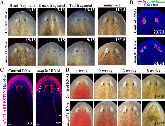
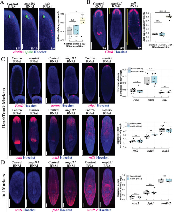
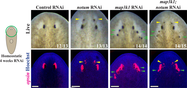
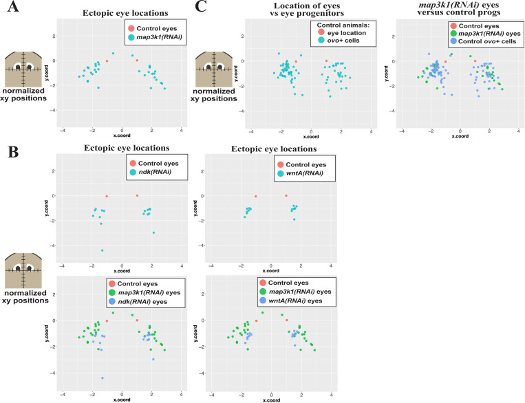
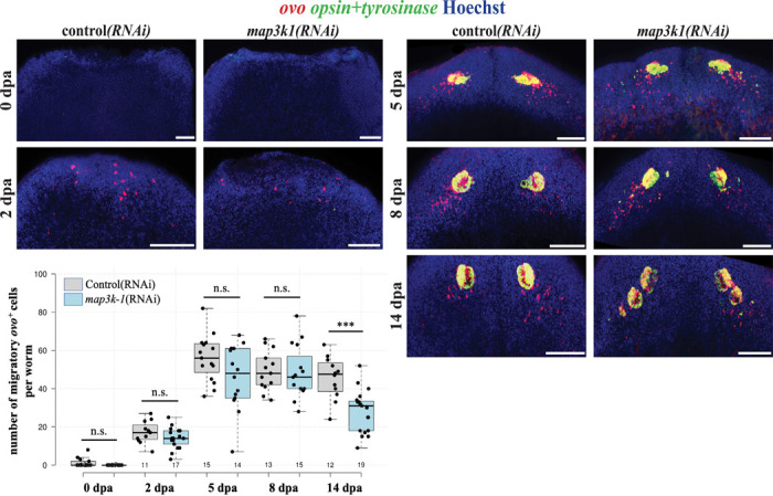
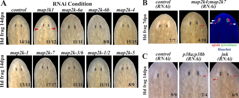
Proper stem cell targeting and differentiation is necessary for regeneration to succeed. In organisms capable of whole body regeneration, considerable progress has been made identifying wound signals initiating this process, but the mechanisms that control the differentiation of progenitors into mature organs are not fully understood. Using the planarian as a model system, we identify a novel function for , a MAP3K family member possessing both kinase and ubiquitin ligase domains, to negatively regulate terminal differentiation of stem cells during eye regeneration. Inhibition of caused the formation of multiple ectopic eyes within the head, but without controlling overall head, brain, or body patterning. By contrast, other known regulators of planarian eye patterning like and also regulate head regionalization, suggesting acts distinctly. Eye resection and regeneration experiments suggest that unlike Wnt signaling perturbation, inhibition did not shift the target destination of eye formation in the animal. Instead, ectopic eyes emerge in the regions normally occupied by migratory eye progenitors, and the onset of ectopic eyes after inhibition coincides with a reduction to eye progenitor numbers. Furthermore, RNAi dosing experiments indicate that progenitors closer to their normal target are relatively more sensitive to the effects of , implicating this factors in controlling the site of terminal differentiation. Eye phenotypes were also observed after inhibition of , and , identifying a putative pathway through which prevents differentiation. Together, these results suggest that regulates a novel control point in the eye regeneration pathway which suppresses the terminal differentiation of progenitors during their migration to target destinations.

摘要

恰当的干细胞靶向和分化是再生成功所必需的。在能够进行全身再生的生物体中,在确定启动这一过程的伤口信号方面已取得了相当大的进展,但控制祖细胞分化为成熟器官的机制尚未完全了解。利用涡虫作为模型系统,我们确定了一个具有激酶和泛素连接酶结构域的MAP3K家族成员的新功能,即在眼睛再生过程中对干细胞的终末分化起负调控作用。抑制该基因导致头部形成多个异位眼,但不影响整体头部、大脑或身体的模式形成。相比之下,其他已知的涡虫眼睛模式形成调节因子,如和,也调节头部区域化,这表明该基因的作用方式不同。眼睛切除和再生实验表明,与Wnt信号扰动不同,抑制该基因并没有改变动物体内眼睛形成的目标位置。相反,异位眼出现在正常情况下由迁移的眼祖细胞占据的区域,抑制该基因后异位眼的出现与眼祖细胞数量的减少同时发生。此外,RNA干扰剂量实验表明,距离其正常目标较近的祖细胞对该基因的影响相对更敏感,这表明该因子在控制终末分化位点方面发挥作用。抑制和后也观察到眼睛表型,从而确定了一条该基因阻止分化的假定途径。总之,这些结果表明,该基因在眼睛再生途径中调节一个新的控制点,在祖细胞迁移到目标位置的过程中抑制其终末分化。

https://cdn.ncbi.nlm.nih.gov/pmc/blobs/032c/11483071/f8a4941dc1b9/nihpp-2024.10.11.617745v1-f0009.jpg
https://cdn.ncbi.nlm.nih.gov/pmc/blobs/032c/11483071/ad5e972decb9/nihpp-2024.10.11.617745v1-f0001.jpg
https://cdn.ncbi.nlm.nih.gov/pmc/blobs/032c/11483071/03d229db8fa9/nihpp-2024.10.11.617745v1-f0002.jpg
https://cdn.ncbi.nlm.nih.gov/pmc/blobs/032c/11483071/7ac5ee4a7220/nihpp-2024.10.11.617745v1-f0003.jpg
https://cdn.ncbi.nlm.nih.gov/pmc/blobs/032c/11483071/57368e8a8bea/nihpp-2024.10.11.617745v1-f0004.jpg
https://cdn.ncbi.nlm.nih.gov/pmc/blobs/032c/11483071/2a3927c4d9fa/nihpp-2024.10.11.617745v1-f0005.jpg
https://cdn.ncbi.nlm.nih.gov/pmc/blobs/032c/11483071/355c808c4186/nihpp-2024.10.11.617745v1-f0006.jpg
https://cdn.ncbi.nlm.nih.gov/pmc/blobs/032c/11483071/4afc264074a6/nihpp-2024.10.11.617745v1-f0007.jpg
https://cdn.ncbi.nlm.nih.gov/pmc/blobs/032c/11483071/56904a560b49/nihpp-2024.10.11.617745v1-f0008.jpg
https://cdn.ncbi.nlm.nih.gov/pmc/blobs/032c/11483071/f8a4941dc1b9/nihpp-2024.10.11.617745v1-f0009.jpg

相似文献

1
suppresses terminal differentiation of migratory eye progenitors in planarian regeneration.抑制涡虫再生过程中迁移性眼祖细胞的终末分化。
bioRxiv. 2024 Oct 12:2024.10.11.617745. doi: 10.1101/2024.10.11.617745.
2
map3k1 suppresses terminal differentiation of migratory eye progenitors in planarian regeneration.Map3k1抑制涡虫再生过程中迁移性眼祖细胞的终末分化。
PLoS Genet. 2025 Mar 17;21(3):e1011457. doi: 10.1371/journal.pgen.1011457. eCollection 2025 Mar.
3
is required for spatial restriction of progenitor differentiation in planarians.涡虫中祖细胞分化的空间限制需要它。
bioRxiv. 2025 Mar 4:2025.03.04.641450. doi: 10.1101/2025.03.04.641450.
4
Src acts with WNT/FGFRL signaling to pattern the planarian anteroposterior axis.Src 通过 WNT/FGFRL 信号作用来模式化扁形动物的前后轴。
Development. 2022 Apr 1;149(7). doi: 10.1242/dev.200125. Epub 2022 Mar 30.
5
Meis1 Controls the Differentiation of Eye Progenitor Cells and the Formation of Posterior Poles during Planarian Regeneration.Meis1在涡虫再生过程中控制眼祖细胞的分化和后极的形成。
Int J Mol Sci. 2023 Feb 9;24(4):3505. doi: 10.3390/ijms24043505.
6
Principles of regeneration revealed by the planarian eye.涡虫眼揭示的再生原则。
Curr Opin Cell Biol. 2021 Dec;73:19-25. doi: 10.1016/j.ceb.2021.05.001. Epub 2021 Jun 13.
7
Wnt/Notum spatial feedback inhibition controls neoblast differentiation to regulate reversible growth of the planarian brain.Wnt/Notum空间反馈抑制控制新生细胞分化以调节涡虫大脑的可逆生长。
Development. 2015 Dec 15;142(24):4217-29. doi: 10.1242/dev.123612. Epub 2015 Nov 2.
8
Integrin suppresses neurogenesis and regulates brain tissue assembly in planarian regeneration.整合素抑制涡虫再生过程中的神经发生并调节脑组织组装。
Development. 2017 Mar 1;144(5):784-794. doi: 10.1242/dev.139964. Epub 2017 Jan 26.
9
Smed-egfr-4 is required for planarian eye regeneration.涡虫眼再生需要Smed-egfr-4。
Int J Dev Biol. 2019;63(1-2):9-15. doi: 10.1387/ijdb.180361fc.
10
Self-organization and progenitor targeting generate stable patterns in planarian regeneration.自组织和祖细胞靶向在扁形动物再生中产生稳定的模式。
Science. 2018 Apr 27;360(6387):404-409. doi: 10.1126/science.aap8179. Epub 2018 Mar 15.

本文引用的文献

1
Crosstalk of MAP3K1 and EGFR signaling mediates gene-environment interactions that block developmental tissue closure.MAP3K1 和 EGFR 信号的串扰介导了阻止发育组织闭合的基因-环境相互作用。
J Biol Chem. 2024 Jul;300(7):107486. doi: 10.1016/j.jbc.2024.107486. Epub 2024 Jun 18.
2
A transcription factor atlas of stem cell fate in planarians.转录因子图谱:扁形动物干细胞命运决定。
Cell Rep. 2024 Mar 26;43(3):113843. doi: 10.1016/j.celrep.2024.113843. Epub 2024 Feb 23.
3
Src acts with WNT/FGFRL signaling to pattern the planarian anteroposterior axis.
Src 通过 WNT/FGFRL 信号作用来模式化扁形动物的前后轴。
Development. 2022 Apr 1;149(7). doi: 10.1242/dev.200125. Epub 2022 Mar 30.
4
Genetic Control of MAP3K1 in Eye Development and Sex Differentiation.基因控制 MAP3K1 在眼睛发育和性别分化中的作用。
Cells. 2021 Dec 23;11(1):34. doi: 10.3390/cells11010034.
5
Ongoing repair of migration-coupled DNA damage allows planarian adult stem cells to reach wound sites.持续修复迁移偶联的 DNA 损伤可使涡虫成体干细胞到达创伤部位。
Elife. 2021 Apr 23;10:e63779. doi: 10.7554/eLife.63779.
6
Muscle and neuronal guidepost-like cells facilitate planarian visual system regeneration.肌肉和神经元类引导细胞促进扁形动物视觉系统再生。
Science. 2020 Jun 26;368(6498). doi: 10.1126/science.aba3203.
7
Smed-egfr-4 is required for planarian eye regeneration.涡虫眼再生需要Smed-egfr-4。
Int J Dev Biol. 2019;63(1-2):9-15. doi: 10.1387/ijdb.180361fc.
8
The planarian Schmidtea mediterranea is a new model to study host-pathogen interactions during fungal infections.地中海涡虫是研究真菌感染期间宿主与病原体相互作用的新模型。
Dev Comp Immunol. 2019 Apr;93:18-27. doi: 10.1016/j.dci.2018.12.005. Epub 2018 Dec 17.
9
The Cellular and Molecular Basis for Planarian Regeneration.涡虫再生的细胞和分子基础。
Cell. 2018 Oct 4;175(2):327-345. doi: 10.1016/j.cell.2018.09.021.
10
Coordination between binocular field and spontaneous self-motion specifies the efficiency of planarians' photo-response orientation behavior.双目视野与自发自我运动之间的协调决定了涡虫光反应定向行为的效率。
Commun Biol. 2018 Sep 21;1:148. doi: 10.1038/s42003-018-0151-2. eCollection 2018.