Department of Cardiology, S. Chiara Hospital, Trento, Italy.
Europace. 2012 Jan;14(1):107-11. doi: 10.1093/europace/eur250. Epub 2011 Aug 18.
The aim of this study is to show the feasibility of a biventricular implantable cardioverter-defibrillator [cardiac resynchronization therapy (CRT)-ICD] implantation using an electroanatomic navigation system and a low dose of fluoroscopy. Here four case reports of patients affected by dilated cardiomyopathy, who underwent cardiac resynchronization therapy, are described.
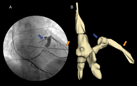
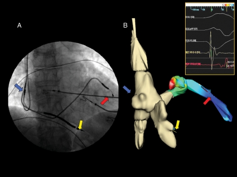
During 2010, four patients were admitted to our Cardiology Department for implantation of an CRT-ICD device in primary prevention. All had an ejection fraction of <35% and were in New York Heart Association class III despite optimal medical therapy. The implantations were performed using the EnSite NavX system. All the leads were positioned in the cardiac chambers utilizing the three-dimensional navigation system and only using X-ray to check that the leads had been positioned correctly. To our knowledge, these cases are the first use of an electroanatomic system for implantation of an CRT-ICD device and in all four cases the cannulation of the coronary sinus (CS) was performed only using the mapping system. Electroanatomic navigation made it possible to minimize X-ray exposure during the implantation of the CRT-ICD device; in addition, the mapping system was used to choose the optimum position of the CS catheter using as reference the maximum activation delay between the two ventricles.
The NavX system shows great potential during the implantation of an CRT-ICD device. It seems to be feasible, safe, and extremely beneficial in terms of a reduction in X-ray exposure. Furthermore, there is benefit of more detailed information and accuracy during the CS lead placement.
本研究旨在展示使用电生理导航系统和低剂量透视进行双心室植入式心脏复律除颤器(心脏再同步治疗除颤器 [CRT-D])植入的可行性。这里描述了 4 例接受心脏再同步治疗的扩张型心肌病患者的病例。
2010 年期间,4 名患者因原发性预防而被收入我院心内科植入 CRT-D 装置。所有患者的射血分数均<35%,尽管接受了最佳药物治疗,但仍处于纽约心脏协会(NYHA)III 级。植入使用 EnSite NavX 系统进行。所有导联均使用三维导航系统定位于心腔,仅使用 X 射线检查以确认导联位置正确。据我们所知,这些病例是首次使用电生理系统植入 CRT-D 装置,在所有 4 例中,冠状窦(CS)的插管仅使用映射系统完成。电生理导航使得在植入 CRT-D 装置期间尽可能减少 X 射线暴露成为可能;此外,映射系统用于使用两个心室之间的最大激活延迟作为参考来选择 CS 导管的最佳位置。
NavX 系统在 CRT-D 装置植入过程中显示出巨大的潜力。它似乎是可行、安全的,并且在减少 X 射线暴露方面具有极大的益处。此外,CS 导联放置的信息更详细、准确性更高。