Vazir Ali, Douthwaite Harriet
Royal Brompton Hospital, National Heart and Lung Institute, Imperial College London, Sydney Street, London SW3 6NP, UK.
J Med Case Rep. 2011 Aug 25;5:417. doi: 10.1186/1752-1947-5-417.
Left atrial myxomas are rare benign tumors of the heart. They vary widely in size, and very little is known about their growth rate. The reported growth rates of left atrial myxomas from several published case reports appears to vary from no growth, to between 1.3 to 6.9 mm/month in diameter within patients with established myxoma who have not undergone surgery.
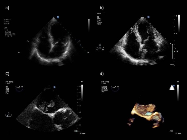
We present the case of a rapidly growing pedunculated left atrial myxoma in a 62-year-old asymptomatic Caucasian woman found incidentally during routine transthoracic echocardiography. Our patient was attending her annual valve clinic assessment for moderate aortic regurgitation, and her two previous consecutive transthoracic echocardiography scans performed 12 and 24 months prior to this appointment had demonstrated a clear left atrium and aortic regurgitation of moderate severity.
To the best of our knowledge, our case is the first to provide images of absence and presence of myxoma from transthoracic echocardiography scans taken a year apart, with estimated growth rate of 2.2 mm/month. Rapidly growing myxoma may be mistaken for thrombus, and may require urgent surgical excision to reduce the risk of associated complications such as thrombo-embolic events, sudden cardiac death and removal of a possibly malignant tumor. The potential for rapid growth should be considered if there is a plan to delay surgery. Furthermore, it would be pertinent to consider annual echocardiography in patients presenting with clinical features suggestive of cardiac myxoma such as constitutional symptoms, as these tumors may be rapid growing and may only become apparent on subsequent echocardiography.
左心房黏液瘤是罕见的心脏良性肿瘤。其大小差异很大,关于其生长速度了解甚少。从几篇已发表的病例报告中可知,未接受手术的确诊左心房黏液瘤患者,其报告的生长速度似乎各不相同,从无生长到直径每月增长1.3至6.9毫米不等。
我们报告一例62岁无症状白种女性的带蒂左心房黏液瘤快速生长病例,该病例在常规经胸超声心动图检查时偶然发现。我们的患者因中度主动脉瓣反流前往年度瓣膜门诊进行评估,在此次就诊前12个月和24个月进行的两次连续经胸超声心动图扫描均显示左心房清晰,有中度严重程度的主动脉瓣反流。
据我们所知,我们的病例是首例提供相隔一年的经胸超声心动图扫描中黏液瘤有无及存在情况图像的病例,估计生长速度为每月2.2毫米。快速生长的黏液瘤可能被误诊为血栓,可能需要紧急手术切除以降低相关并发症的风险,如血栓栓塞事件、心源性猝死以及切除可能恶变的肿瘤。如果计划延迟手术,应考虑到其快速生长的可能性。此外,对于出现提示心脏黏液瘤临床特征(如全身症状)的患者,应考虑每年进行超声心动图检查,因为这些肿瘤可能生长迅速,可能仅在后续超声心动图检查中才会显现。