Laboratory of Molecular Neurobiology & Biophysics, The Rockefeller University, Howard Hughes Medical Institute, 1230 York Avenue, New York, New York 10065, USA.
Nature. 2011 Aug 28;477(7365):495-8. doi: 10.1038/nature10370.
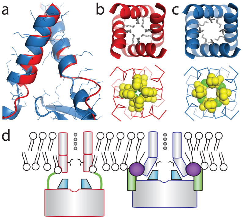
The regulation of ion channel activity by specific lipid molecules is widely recognized as an integral component of electrical signalling in cells. In particular, phosphatidylinositol 4,5-bisphosphate (PIP(2)), a minor yet dynamic phospholipid component of cell membranes, is known to regulate many different ion channels. PIP(2) is the primary agonist for classical inward rectifier (Kir2) channels, through which this lipid can regulate a cell's resting membrane potential. However, the molecular mechanism by which PIP(2) exerts its action is unknown. Here we present the X-ray crystal structure of a Kir2.2 channel in complex with a short-chain (dioctanoyl) derivative of PIP(2). We found that PIP(2) binds at an interface between the transmembrane domain (TMD) and the cytoplasmic domain (CTD). The PIP(2)-binding site consists of a conserved non-specific phospholipid-binding region in the TMD and a specific phosphatidylinositol-binding region in the CTD. On PIP(2) binding, a flexible expansion linker contracts to a compact helical structure, the CTD translates 6 Å and becomes tethered to the TMD and the inner helix gate begins to open. In contrast, the small anionic lipid dioctanoyl glycerol pyrophosphatidic acid (PPA) also binds to the non-specific TMD region, but not to the specific phosphatidylinositol region, and thus fails to engage the CTD or open the channel. Our results show how PIP(2) can control the resting membrane potential through a specific ion-channel-receptor-ligand interaction that brings about a large conformational change, analogous to neurotransmitter activation of ion channels at synapses.
特定脂质分子对离子通道活性的调节被广泛认为是细胞电信号传递的一个组成部分。特别是磷脂酰肌醇 4,5-二磷酸(PIP(2)),作为细胞膜中一种少量但动态的磷脂成分,已知可调节许多不同的离子通道。PIP(2)是经典内向整流(Kir2)通道的主要激动剂,通过这种脂质可以调节细胞的静息膜电位。然而,PIP(2)发挥作用的分子机制尚不清楚。在这里,我们展示了一个 Kir2.2 通道与 PIP(2)的短链(二辛酰)衍生物复合物的 X 射线晶体结构。我们发现 PIP(2)结合在跨膜域(TMD)和细胞质域(CTD)之间的界面上。PIP(2)结合位点由 TMD 中保守的非特异性磷脂结合区和 CTD 中特异性的磷脂酰肌醇结合区组成。在 PIP(2)结合时,一个灵活的扩展接头收缩到一个紧凑的螺旋结构,CTD 平移 6 Å 并与 TMD 连接,内螺旋门开始打开。相比之下,小分子阴离子脂质二辛酰甘油焦磷酸脂酸(PPA)也结合到非特异性 TMD 区域,但不结合特异性的磷脂酰肌醇区域,因此无法与 CTD 结合或打开通道。我们的结果表明,PIP(2)如何通过一种特定的离子通道-受体-配体相互作用来控制静息膜电位,这种相互作用引起了很大的构象变化,类似于神经递质在突触处激活离子通道。