Departamento de Microbiologia e Imunologia, UNESP - Univ Estadual Paulista, Instituto de Biociências - Campus Botucatu, CEP 18618-970, SP, Brasil.
J Inflamm (Lond). 2011 Aug 29;8:23. doi: 10.1186/1476-9255-8-23.
Arterial peripheral disease is a condition caused by the blocked blood flow resulting from arterial cholesterol deposits within the arms, legs and aorta. Studies have shown that macrophages in atherosclerotic plaque are highly activated, which makes these cells important antigen-presenting cells that develop a specific immune response, in which LDLox is the inducing antigen. As functional changes of cells which participate in the atherogenesis process may occur in the peripheral blood, the objectives of the present study were to evaluate plasma levels of anti-inflammatory and inflammatory cytokines including TNF-α, IFN-γ, interleukin-6 (IL-6), IL-10 and TGF-β in patients with peripheral arteriosclerosis obliterans, to assess the monocyte activation level in peripheral blood through the ability of these cells to release hydrogen peroxide (H2O2) and to develop fungicidal activity against Candida albicans (C. albicans) in vitro.
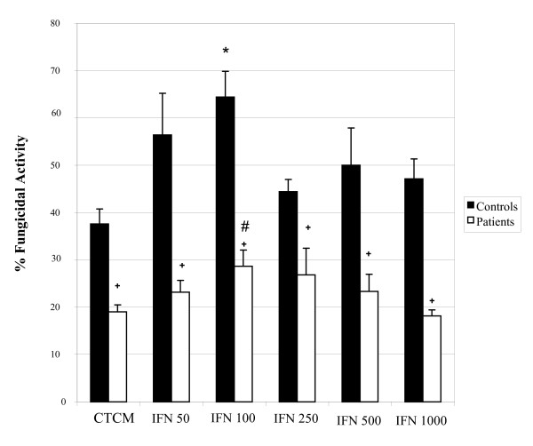
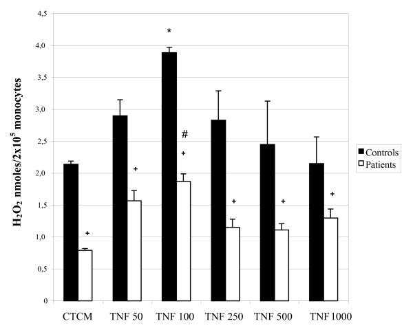
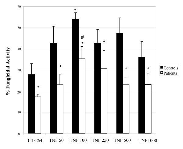
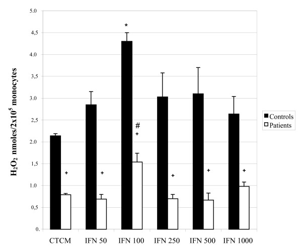
TNF-α, IFN-γ, IL-6, IL-10 and TGF-β from plasma of patients were detected by ELISA. Monocyte cultures activated in vitro with TNF-alpha and IFN-gamma were evaluated by fungicidal activity against C. albicans by culture plating and Colony Forming Unit (CFU) recovery, and by H2O2 production.
Plasma levels of all cytokines were significantly higher in patients compared to those detected in control subjects. Control group monocytes did not release substantial levels of H2O2 in vitro, but these levels were significantly increased after activation with IFN-γ and TNF-α. Monocytes of patients, before and after activation, responded less than those of control subjects. Similar results were found when fungicidal activity was evaluated. The results seen in patients were always significantly smaller than among control subjects.
The results revealed an unresponsiveness of patient monocytes in vitro probably due to the high activation process occurring in vivo as corroborated by high plasma cytokine levels.
周围动脉疾病是一种由手臂、腿部和主动脉内动脉胆固醇沉积导致血流阻塞引起的疾病。研究表明,动脉粥样硬化斑块中的巨噬细胞高度激活,这使得这些细胞成为重要的抗原提呈细胞,引发特定的免疫反应,其中 LDLox 是诱导抗原。由于参与动脉粥样硬化形成过程的细胞可能会发生功能变化,因此本研究旨在评估周围动脉硬化闭塞症患者的抗炎和促炎细胞因子(包括 TNF-α、IFN-γ、白细胞介素-6 [IL-6]、IL-10 和 TGF-β)的血浆水平,通过这些细胞释放过氧化氢 (H2O2) 的能力来评估外周血单核细胞的激活水平,并评估其体外对白色念珠菌 (C. albicans) 的杀菌活性。
通过 ELISA 检测患者血浆中的 TNF-α、IFN-γ、IL-6、IL-10 和 TGF-β。通过培养平板和集落形成单位 (CFU) 回收以及 H2O2 产生,评估体外用 TNF-α和 IFN-γ激活的单核细胞对 C. albicans 的杀菌活性。
与对照组相比,患者的所有细胞因子的血浆水平均显着升高。对照组单核细胞在体外没有释放大量的 H2O2,但在激活后 IFN-γ和 TNF-α的水平显着增加。患者的单核细胞,无论是在激活前还是激活后,反应都低于对照组。当评估杀菌活性时,也发现了类似的结果。患者的结果总是明显小于对照组。
结果显示患者的单核细胞在体外无反应性,可能是由于体内高激活过程所致,这与高细胞因子水平相符。