Cancer Biology Program, University of Pennsylvania, Philadelphia, Pennsylvania, USA.
J Cell Physiol. 2012 Jun;227(6):2654-9. doi: 10.1002/jcp.23007.
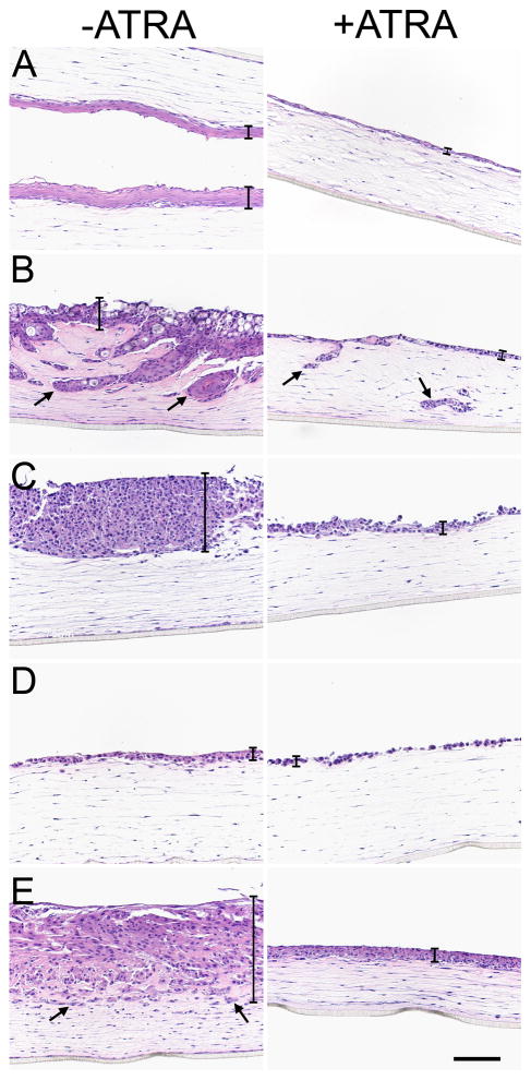
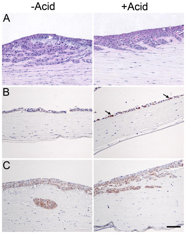
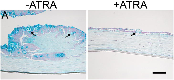
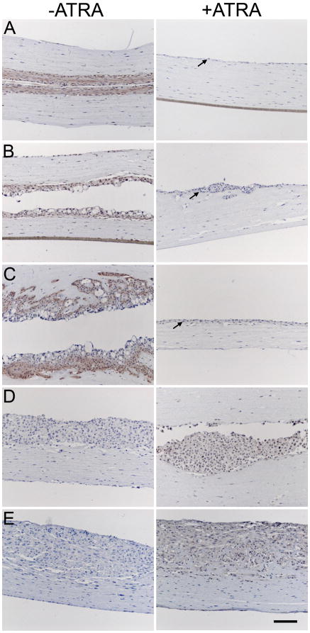
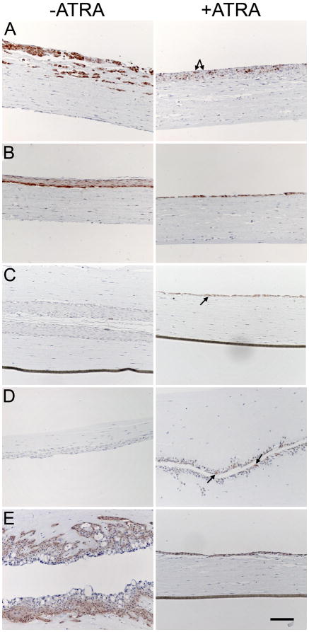
Understanding the molecular and cellular processes underlying the development, maintenance, and progression of Barrett's esophagus (BE) presents an empirical challenge because there are no simple animal models and standard 2D cell culture can distort cellular processes. Here we describe a three-dimensional (3D) cell culture system to study BE. BE cell lines (CP-A, CP-B, CP-C, and CP-D) and esophageal squamous keratinocytes (EPC2) were cultured on a matrix consisting of esophageal fibroblasts and collagen. Comparison of growth and cytokeratin expression in the presence of all-trans retinoic acid or hydrochloric acid was made by immunohistochemistry and Alcian Blue staining to determine which treatments produced a BE phenotype of columnar cytokeratin expression in 3D culture. All-trans retinoic acid differentially affected the growth of BE cell lines in 3D culture. Notably, the non-dyplastic metaplasia-derived cell line (CP-A) expressed reduced squamous cytokeratins and enhanced columnar cytokeratins upon ATRA treatment. ATRA altered the EPC2 squamous cytokeratin profile towards a more columnar expression pattern. Cell lines derived from patients with high-grade dysplasia already expressed columnar cytokeratins and therefore did not show a systematic shift toward a more columnar phenotype with ATRA treatment. ATRA treatment, however, did reduce the squamoid-like multilayer stratification observed in all cell lines. As the first study to demonstrate long-term 3D growth of BE cell lines, we have determined that BE cells can be cultured for at least 3 weeks on a fibroblast/collagen matrix and that the use of ATRA causes a general reduction in squamous-like multilayered growth and an increase in columnar phenotype with the specific effects cell-line dependent.
了解巴雷特食管 (BE) 发展、维持和进展的分子和细胞过程带来了实证挑战,因为没有简单的动物模型,标准的 2D 细胞培养可能会扭曲细胞过程。在这里,我们描述了一种用于研究 BE 的三维 (3D) 细胞培养系统。BE 细胞系(CP-A、CP-B、CP-C 和 CP-D)和食管鳞状角质形成细胞(EPC2)在由食管成纤维细胞和胶原蛋白组成的基质上培养。通过免疫组织化学和阿利新蓝染色比较存在全反式视黄酸或盐酸时的生长和细胞角蛋白表达,以确定哪种处理在 3D 培养中产生 BE 柱状细胞角蛋白表达的表型。全反式视黄酸对 3D 培养中 BE 细胞系的生长有不同的影响。值得注意的是,非异型增生性化生衍生的细胞系(CP-A)在 ATRA 处理后表达减少的鳞状细胞角蛋白,并增强柱状细胞角蛋白。ATRA 改变了 EPC2 鳞状细胞角蛋白的表达模式,向更柱状的表达模式转变。来自高级别异型增生患者的细胞系已经表达柱状细胞角蛋白,因此在用 ATRA 处理时不会表现出向更柱状表型的系统转变。然而,ATRA 处理确实减少了所有细胞系中观察到的鳞状样多层分层。作为首次证明 BE 细胞系长期 3D 生长的研究,我们已经确定 BE 细胞可以在成纤维细胞/胶原蛋白基质上培养至少 3 周,并且 ATRA 的使用导致鳞状样多层生长普遍减少,柱状表型增加,具体影响取决于细胞系。