Faculty of Medicine, Institute of Cell Biology, University of Ljubljana, Ljubljana, Slovenia.
PLoS One. 2011;6(8):e23636. doi: 10.1371/journal.pone.0023636. Epub 2011 Aug 24.
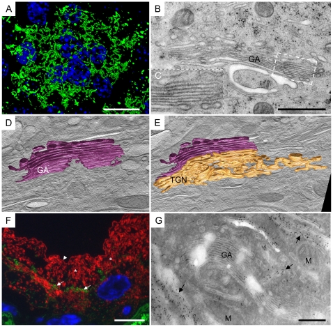
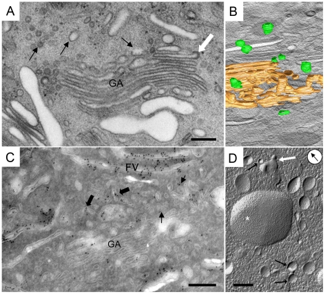
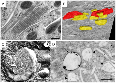
Urothelial plaques are specialized membrane domains in urothelial superficial (umbrella) cells, composed of highly ordered uroplakin particles. We investigated membrane compartments involved in the formation of urothelial plaques in mouse umbrella cells. The Golgi apparatus did not contain uroplakins organized into plaques. In the post-Golgi region, three distinct membrane compartments containing uroplakins were characterized: i) Small rounded vesicles, located close to the Golgi apparatus, were labelled weakly with anti-uroplakin antibodies and they possessed no plaques; we termed them "uroplakin-positive transporting vesicles" (UPTVs). ii) Spherical-to-flattened vesicles, termed "immature fusiform vesicles" (iFVs), were uroplakin-positive in their central regions and contained small urothelial plaques. iii) Flattened "mature fusiform vesicles" (mFVs) contained large plaques, which were densely labelled with anti-uroplakin antibodies. Endoytotic marker horseradish peroxidase was not found in these post-Golgi compartments. We propose a detailed model of de novo urothelial plaque formation in post-Golgi compartments: UPTVs carrying individual 16-nm particles detach from the Golgi apparatus and subsequently fuse into iFV. Concentration of 16-nm particles into plaques and removal of uroplakin-negative membranes takes place in iFVs. With additional fusions and buddings, iFVs mature into mFVs, each carrying two urothelial plaques toward the apical surface of the umbrella cell.
尿路上皮斑是尿路上皮浅层(伞状)细胞中特化的膜域,由高度有序的尿路上皮蛋白颗粒组成。我们研究了参与小鼠伞状细胞尿路上皮斑形成的膜隔室。高尔基氏体中不含有组织成斑的尿路上皮蛋白。在后高尔基区,有三个含有尿路上皮蛋白的不同膜隔室被鉴定为:i)靠近高尔基氏体的小圆形囊泡,用抗尿路上皮蛋白抗体弱标记,并且没有斑,我们将其称为“尿路上皮蛋白阳性转运囊泡”(UPTVs)。ii)球形至扁平的囊泡,称为“未成熟梭形囊泡”(iFVs),在中央区域呈尿路上皮蛋白阳性,并含有小的尿路上皮斑。iii)扁平的“成熟梭形囊泡”(mFVs)含有大的斑,这些斑与抗尿路上皮蛋白抗体强烈标记。内吞标记物辣根过氧化物酶未在这些高尔基后隔室中发现。我们提出了高尔基后隔室中尿路上皮斑形成的详细模型:携带单个 16nm 颗粒的 UPTVs 从高尔基氏体上脱离,随后融合成 iFV。16nm 颗粒在 iFVs 中浓缩成斑,并去除尿路上皮蛋白阴性的膜。随着进一步的融合和芽生,iFVs 成熟为 mFVs,每个 mFVs 向伞状细胞的顶端表面携带两个尿路上皮斑。