Habermehl Christina, Schmitz Christoph H, Steinbrink Jens
Berlin NeuroImaging Center, Charité University Hospital, Department of Neurology, Berlin, Germany.
Opt Express. 2011 Sep 12;19(19):18636-44. doi: 10.1364/OE.19.018636.
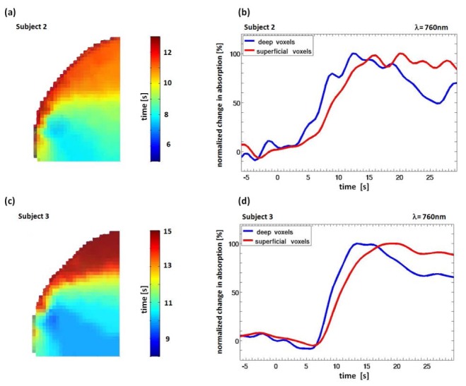
Non-invasive diffuse optical tomography (DOT) of the adult brain has recently been shown to improve the spatial resolution for functional brain imaging applications. Here we show that high-resolution (HR) DOT is also advantageous for clinical perfusion imaging using an optical contrast agent. We present the first HR-DOT results with a continuous wave near infrared spectroscopy setup using a dense grid of optical fibers and indocyanine green (ICG) as an exogenic contrast agent. We find an early arrival of the ICG bolus in the intracerebral tissue and a delayed arrival of the bolus in the extracerebral tissue, achieving the separation of both layers. This demonstrates the method's potential for brain perfusion monitoring in neurointensive care patients.
成人脑部的无创漫射光学断层扫描(DOT)最近已被证明可提高功能性脑成像应用的空间分辨率。在此,我们表明高分辨率(HR)DOT对于使用光学造影剂的临床灌注成像也具有优势。我们展示了使用密集光纤网格和吲哚菁绿(ICG)作为外源性造影剂的连续波近红外光谱设置下的首个HR-DOT结果。我们发现ICG团注在脑组织中提前到达,而在脑组织外延迟到达,实现了两层的分离。这证明了该方法在神经重症监护患者脑灌注监测中的潜力。