Chamié Daniel, Wang Zhao, Bezerra Hiram, Rollins Andrew M, Costa Marco A
Curr Cardiovasc Imaging Rep. 2011 Aug;4(4):276-283. doi: 10.1007/s12410-011-9090-8. Epub 2011 May 12.
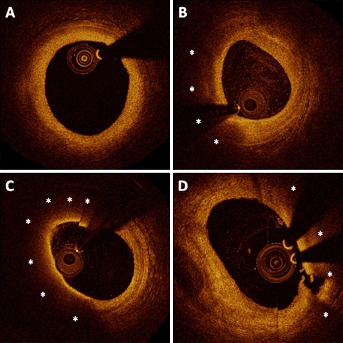
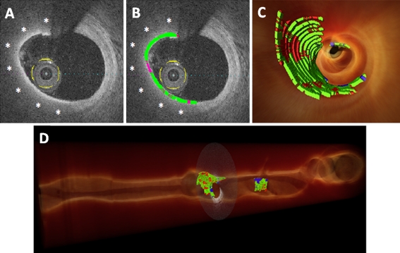
The pathophysiology of acute coronary syndromes has long been associated with atherosclerotic plaque rupture. Inflammation, thinning, and disruption of the fibrous cap have been implicated with the final processes leading to plaque rupture, but confirmation of these mechanisms of coronary thrombosis in humans has been hampered by the lack of imaging methods with sufficient resolution to resolve fibrous cap characterization and thickness in vivo. Intravascular optical coherence tomography (OCT) provides images with micron-level axial and lateral resolution, enabling detailed visualization of micro-structural changes of the arterial wall. The present article provides an overview of the potential role of OCT in identifying and characterizing fibrous cap morphology, thickness, and inflammation in human coronary plaques.
急性冠状动脉综合征的病理生理学长期以来一直与动脉粥样硬化斑块破裂相关。纤维帽的炎症、变薄和破裂与导致斑块破裂的最终过程有关,但由于缺乏具有足够分辨率以在体内分辨纤维帽特征和厚度的成像方法,人类冠状动脉血栓形成的这些机制尚未得到证实。血管内光学相干断层扫描(OCT)提供具有微米级轴向和横向分辨率的图像,能够详细可视化动脉壁的微观结构变化。本文概述了OCT在识别和表征人类冠状动脉斑块中纤维帽形态、厚度及炎症方面的潜在作用。