Soliman Osama I I, Anwar Ashraf M, Metawei Ahmed K, McGhie Jackie S, Geleijnse Marcel L, Ten Cate Folkert J
Curr Cardiovasc Imaging Rep. 2011 Oct;4(5):370-377. doi: 10.1007/s12410-011-9099-z. Epub 2011 Jul 9.
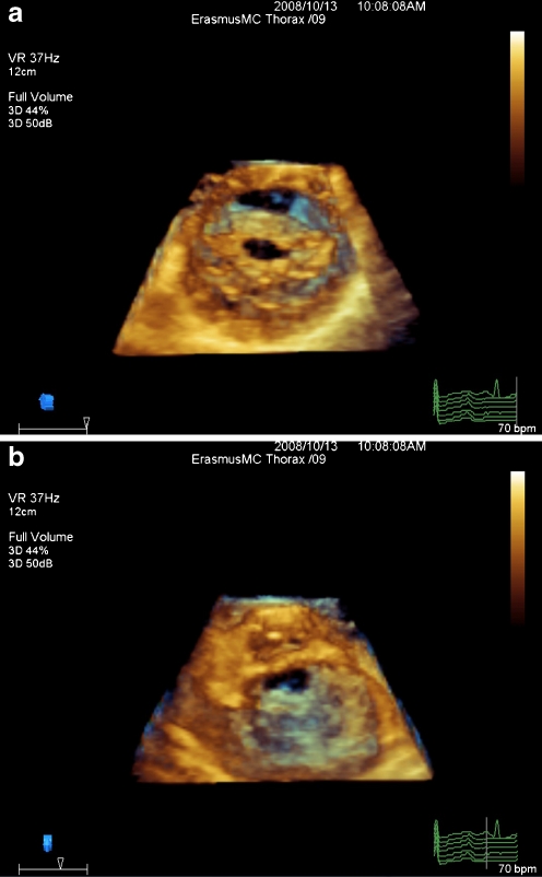
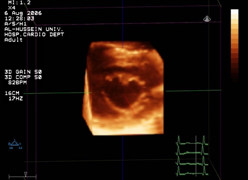
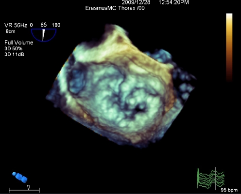
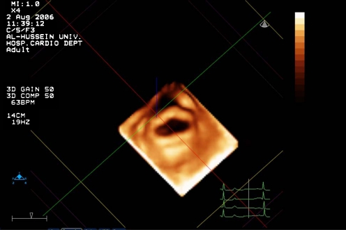
Nonsurgical management of patients with symptomatic mitral valve stenosis has been established as the therapeutic modality of choice for two decades. Catheter-based balloon dilation of the stenotic valvular area has been shown, at least, as effective as surgical interventions. Unfavorable results of catheter-based interventions are largely due to unfavorable morphology of the valve apparatus, particularly leaflets calcification and subvalvular apparatus involvement. A mitral valve score has been proposed in Boston, MA, about two decades ago, based on morphologic assessment of mitral valve apparatus by two-dimensional (2D) echocardiography to predict successful balloon dilation of the mitral valve. Several other scores have been developed in the following years in order to more successfully predict balloon dilatation outcome. However, all those scores were based on 2D echocardiography, which is limited by ability to distinguish calcification and subvalvular involvement. The introduction of new matrix-based ultrasound probe has allowed 3D echocardiography (3DE) to provide more detailed morphologic analysis of mitral valve apparatus including calcification and subvalvular involvement. Recently, a new 3DE scoring system has been proposed by our group, which represents an important leap into refinement of the use of echocardiography guiding mitral valve interventions.
二十年来,有症状的二尖瓣狭窄患者的非手术治疗已被确立为首选治疗方式。经导管球囊扩张狭窄瓣膜区域已被证明至少与手术干预一样有效。经导管干预的不良结果主要归因于瓣膜装置的不良形态,尤其是瓣叶钙化和瓣下装置受累。大约二十年前,在马萨诸塞州波士顿提出了二尖瓣评分,基于二维(2D)超声心动图对二尖瓣装置的形态学评估来预测二尖瓣球囊扩张的成功率。在接下来的几年里,又开发了其他几种评分系统,以便更成功地预测球囊扩张结果。然而,所有这些评分都是基于二维超声心动图,其在区分钙化和瓣下受累方面存在局限性。新型基于矩阵的超声探头的引入使三维超声心动图(3DE)能够对二尖瓣装置进行更详细的形态学分析,包括钙化和瓣下受累情况。最近,我们团队提出了一种新的三维超声心动图评分系统,这代表了在利用超声心动图指导二尖瓣干预方面的重要飞跃。