Embryo Implantation Laboratory, Prince Henry's Institute, Clayton, Melbourne, Australia.
PLoS One. 2011;6(9):e25288. doi: 10.1371/journal.pone.0025288. Epub 2011 Sep 23.
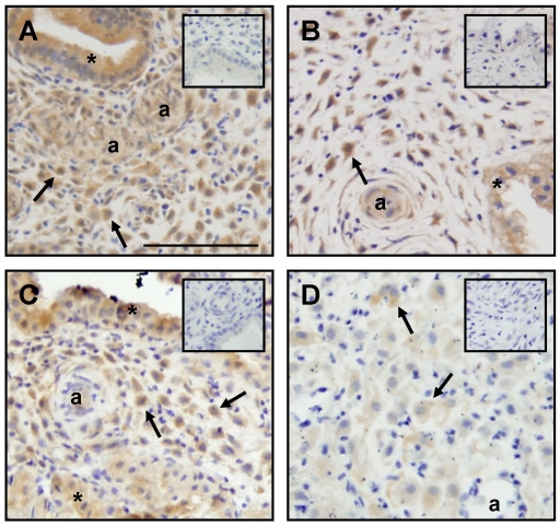
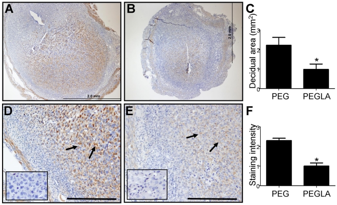
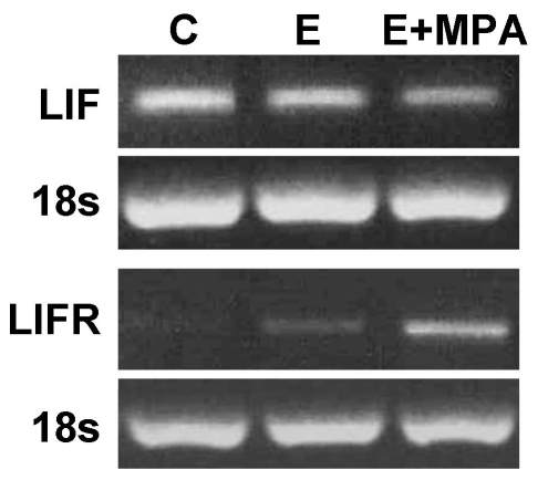
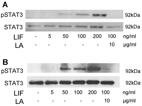
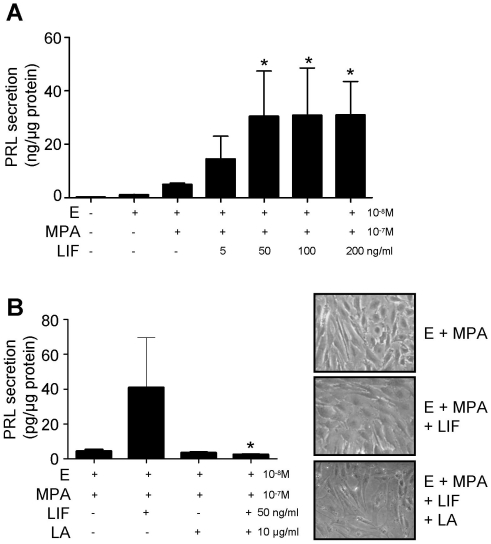
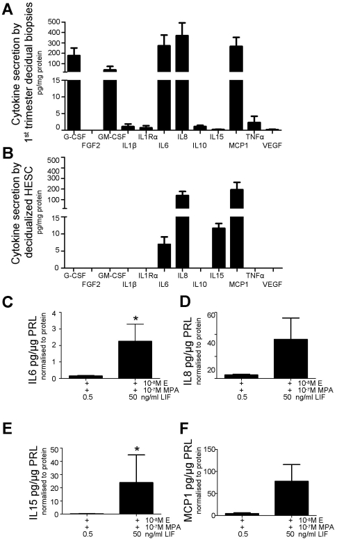
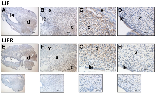
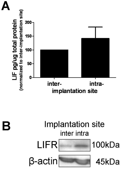
Adequate differentiation or decidualization of endometrial stromal cells (ESC) is critical for successful pregnancy in humans and rodents. Here, we investigated the role of leukemia inhibitory factor (LIF) in human and murine decidualization. Ex vivo human (H) ESC decidualization was induced by estrogen (E, 10(-8) M) plus medroxyprogesterone acetate (MPA, 10(-7) M). Exogenous LIF (≥50 ng/ml) induced STAT3 phosphorylation in non-decidualized and decidualized HESC and enhanced E+MPA-induced decidualization (measured by PRL secretion, P<0.05). LIF mRNA in HESC was down-regulated by decidualization treatment (E+MPA) whereas LIF receptor (R) mRNA was up-regulated, suggesting that the decidualization stimulus 'primed' HESC for LIF action, but that factors not present in our in vitro model were required to induce LIF expression. Ex vivo first trimester decidual biopsies secreted >100 pg/mg G-CSF, IL6, IL8, and MCP1. Decidualized HESC secreted IL6, IL8, IL15 and MCP1. LIF (50 ng/ml) up-regulated IL6 and IL15 (P<0.05) secretion in decidualized HESC compared to 0.5 ng/ml LIF. In murine endometrium, LIF and LIFR immunolocalized to decidualized stromal cells on day 5 of gestation (day 0 = day of plug detection). Western blotting confirmed that LIF and the LIFR were up-regulated in intra-implantation sites compared to inter-implantation sites on Day 5 of gestation. To determine the role of LIF during in vivo murine decidualization, intra-peritoneal injections of a long-acting LIF antagonist (PEGLA; 900 or 1200 µg) were given just post-attachment, during the initiation of decidualization on day 4. PEGLA treatment reduced implantation site decidual area (P<0.05) and desmin staining immuno-intensity (P<0.05) compared to control on day 6 of gestation. This study demonstrated that LIF was an important regulator of decidualization in humans and mice and data provides insight into the processes underlying decidualization, which are important for understanding implantation and placentation.
子宫内膜基质细胞(ESC)的充分分化或蜕膜化对于人类和啮齿动物的成功妊娠至关重要。在这里,我们研究了白血病抑制因子(LIF)在人类和鼠类蜕膜化中的作用。通过雌激素(E,10(-8)M)加醋酸甲羟孕酮(MPA,10(-7)M)诱导离体人(H)ESC 蜕膜化。外源性 LIF(≥50ng/ml)诱导未蜕膜化和蜕膜化的 HESC 中 STAT3 磷酸化,并增强 E+MPA 诱导的蜕膜化(通过 PRL 分泌测量,P<0.05)。蜕膜化处理(E+MPA)下调 HESC 中的 LIFmRNA,而 LIF 受体(R)mRNA 上调,这表明蜕膜化刺激物“启动”HESC 进行 LIF 作用,但我们的体外模型中不存在诱导 LIF 表达所需的因素。离体早期妊娠蜕膜活检分泌>100pg/mg G-CSF、IL6、IL8 和 MCP1。蜕膜化的 HESC 分泌 IL6、IL8、IL15 和 MCP1。与 0.5ng/ml LIF 相比,50ng/ml LIF 上调蜕膜化 HESC 中的 IL6 和 IL15 分泌(P<0.05)。在鼠类子宫内膜中,LIF 和 LIFR 免疫定位在妊娠第 5 天的蜕膜化基质细胞上(0 天=栓检测日)。Western 印迹证实,与妊娠第 5 天的植入间隔相比,植入部位的 LIF 和 LIFR 在妊娠第 5 天的植入部位上调。为了确定 LIF 在体内鼠类蜕膜化过程中的作用,在第 4 天蜕膜化开始时,即在附着后立即给予腹膜内注射长效 LIF 拮抗剂(PEGLA;900 或 1200μg)。与妊娠第 6 天的对照相比,PEGLA 处理减少了植入部位蜕膜面积(P<0.05)和结蛋白染色免疫强度(P<0.05)。本研究表明,LIF 是人类和小鼠蜕膜化的重要调节剂,数据为了解蜕膜化的过程提供了线索,这些过程对于理解植入和胎盘形成很重要。