Department of Molecular Cell Biology, Section Electron Microscopy, Leiden University Medical Center, Leiden, The Netherlands.
mBio. 2011 Oct 4;2(5). doi: 10.1128/mBio.00166-11. Print 2011.
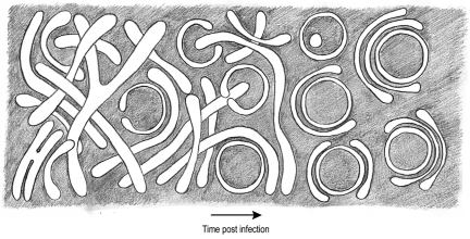
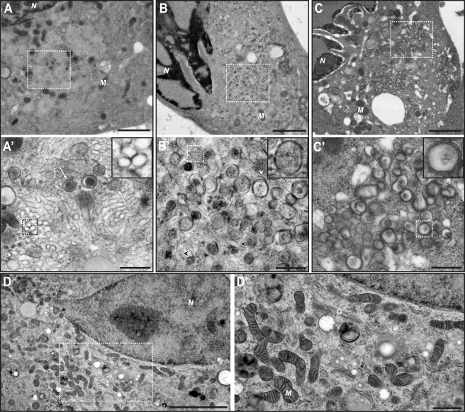
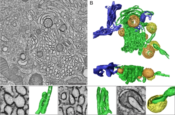
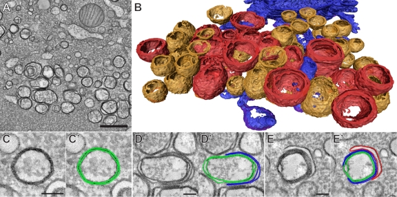
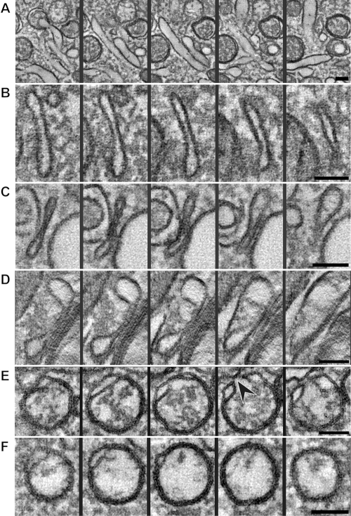
All positive-strand RNA viruses induce membrane structures in their host cells which are thought to serve as suitable microenvironments for viral RNA synthesis. The structures induced by enteroviruses, which are members of the family Picornaviridae, have so far been described as either single- or double-membrane vesicles (DMVs). Aside from the number of delimiting membranes, their exact architecture has also remained elusive due to the limitations of conventional electron microscopy. In this study, we used electron tomography (ET) to solve the three-dimensional (3-D) ultrastructure of these compartments. At different time points postinfection, coxsackievirus B3-infected cells were high-pressure frozen and freeze-substituted for ET analysis. The tomograms showed that during the exponential phase of viral RNA synthesis, closed smooth single-membrane tubules constituted the predominant virus-induced membrane structure, with a minor proportion of DMVs that were either closed or connected to the cytosol in a vase-like configuration. As infection progressed, the DMV number steadily increased, while the tubular single-membrane structures gradually disappeared. Late in infection, complex multilamellar structures, previously unreported, became apparent in the cytoplasm. Serial tomography disclosed that their basic unit is a DMV, which is enwrapped by one or multiple cisternae. ET also revealed striking intermediate structures that strongly support the conversion of single-membrane tubules into double-membrane and multilamellar structures by a process of membrane apposition, enwrapping, and fusion. Collectively, our work unravels the sequential appearance of distinct enterovirus-induced replication structures, elucidates their detailed 3-D architecture, and provides the basis for a model for their transformation during the course of infection.
Positive-strand RNA viruses hijack specific intracellular membranes and remodel them into special structures that support viral RNA synthesis. The ultrastructural characterization of these "replication structures" is key to understanding their precise role. Here, we resolved the three-dimensional architecture of enterovirus-induced membranous compartments and their transformation in time by applying electron tomography to cells infected with coxsackievirus B3 (CVB3). Our results show that closed single-membrane tubules are the predominant initial virus-induced structure, whereas double-membrane vesicles (DMVs) become increasingly abundant at the expense of these tubules as infection progresses. Additionally, more complex multilamellar structures appear late in infection. Based on compelling intermediate structures in our tomograms, we propose a model for transformation from the tubules to DMVs and multilamellar structures via enwrapping events. Our work provides an in-depth analysis of the development of an unsuspected variety of distinct replication structures during the course of CVB3 infection.
所有正链 RNA 病毒都会在宿主细胞中诱导膜结构,这些结构被认为是病毒 RNA 合成的合适微环境。肠道病毒(属于小 RNA 病毒科)诱导的结构迄今为止被描述为单膜囊泡或双膜囊泡 (DMV)。除了分隔膜的数量外,由于传统电子显微镜的限制,其确切的结构仍然难以捉摸。在这项研究中,我们使用电子断层扫描 (ET) 来解决这些隔室的三维 (3-D) 超微结构。在 Coxsackievirus B3 感染后的不同时间点,细胞进行高压冷冻和冷冻置换以进行 ET 分析。断层扫描显示,在病毒 RNA 合成的指数期,闭合的光滑单膜管构成了主要的病毒诱导膜结构,而 DMV 的比例较小,要么闭合,要么以花瓶状与细胞质相连。随着感染的进展,DMV 的数量稳步增加,而管状单膜结构逐渐消失。在感染后期,细胞质中出现了以前未报道过的复杂的多层结构。连续断层扫描显示,它们的基本单位是一个 DMV,被一个或多个内质网包裹。ET 还揭示了引人注目的中间结构,强烈支持通过膜贴附、包裹和融合过程将单膜管转化为双膜和多层结构。总的来说,我们的工作揭示了不同肠道病毒诱导的复制结构的顺序出现,阐明了它们的详细 3-D 结构,并为感染过程中它们的转化提供了模型基础。
正链 RNA 病毒劫持特定的细胞内膜,并将其重塑为支持病毒 RNA 合成的特殊结构。这些“复制结构”的超微结构特征是理解其精确作用的关键。在这里,我们通过应用电子断层扫描技术对感染柯萨奇病毒 B3 (CVB3) 的细胞进行研究,解析了肠道病毒诱导的膜性隔室的三维结构及其随时间的转化。我们的结果表明,闭合的单膜管是主要的初始病毒诱导结构,而随着感染的进展,双膜囊泡 (DMV) 变得越来越多,这些管逐渐减少。此外,在感染后期会出现更复杂的多层结构。基于我们断层扫描中的有力中间结构,我们提出了一个从管到 DMV 和多层结构的包裹事件的转化模型。我们的工作对 CVB3 感染过程中不同复制结构的发展进行了深入分析。