Knoops Kèvin, Kikkert Marjolein, Worm Sjoerd H E van den, Zevenhoven-Dobbe Jessika C, van der Meer Yvonne, Koster Abraham J, Mommaas A Mieke, Snijder Eric J
Section Electron Microscopy, Department of Molecular Cell Biology, Leiden University Medical Center, Leiden, The Netherlands.
PLoS Biol. 2008 Sep 16;6(9):e226. doi: 10.1371/journal.pbio.0060226.
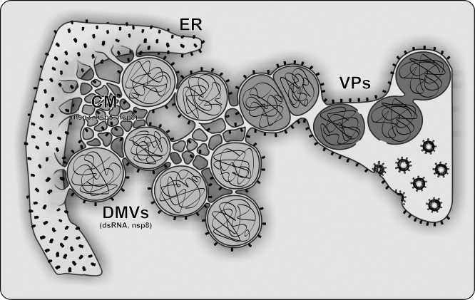
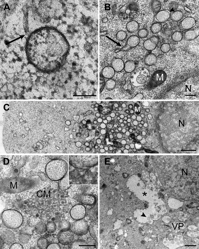
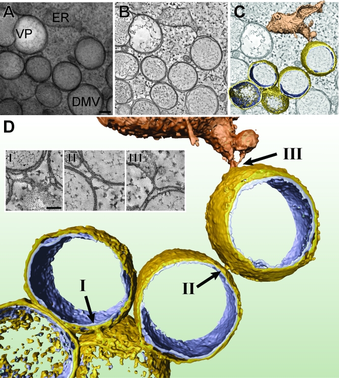
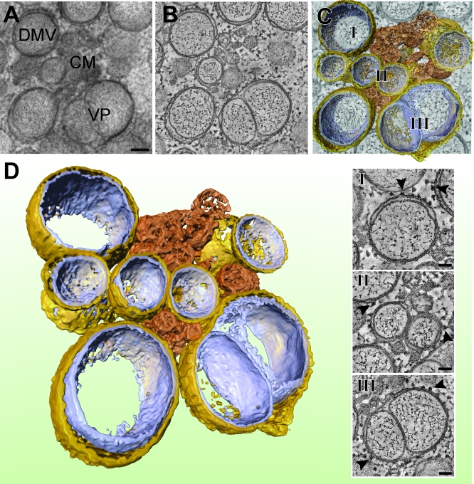
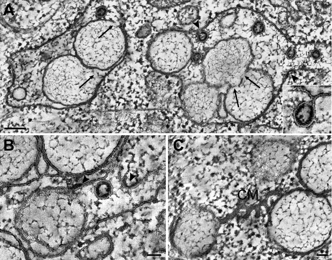
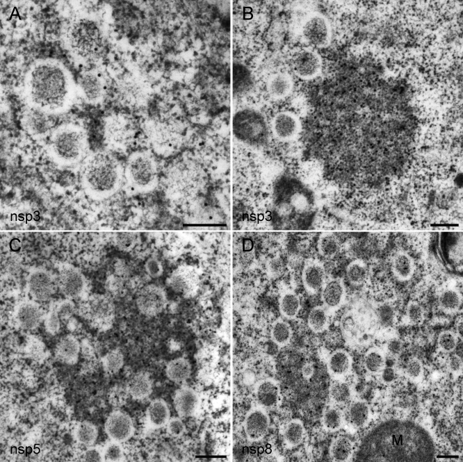
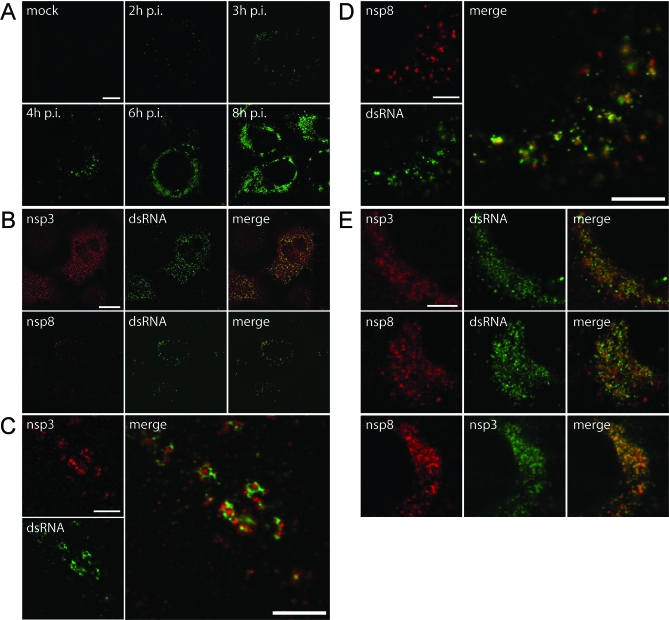
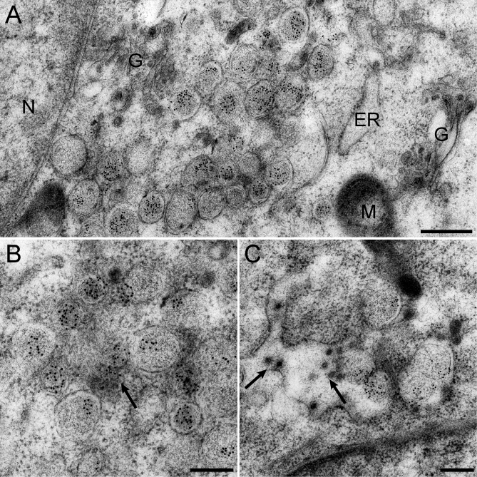
Positive-strand RNA viruses, a large group including human pathogens such as SARS-coronavirus (SARS-CoV), replicate in the cytoplasm of infected host cells. Their replication complexes are commonly associated with modified host cell membranes. Membrane structures supporting viral RNA synthesis range from distinct spherular membrane invaginations to more elaborate webs of packed membranes and vesicles. Generally, their ultrastructure, morphogenesis, and exact role in viral replication remain to be defined. Poorly characterized double-membrane vesicles (DMVs) were previously implicated in SARS-CoV RNA synthesis. We have now applied electron tomography of cryofixed infected cells for the three-dimensional imaging of coronavirus-induced membrane alterations at high resolution. Our analysis defines a unique reticulovesicular network of modified endoplasmic reticulum that integrates convoluted membranes, numerous interconnected DMVs (diameter 200-300 nm), and "vesicle packets" apparently arising from DMV merger. The convoluted membranes were most abundantly immunolabeled for viral replicase subunits. However, double-stranded RNA, presumably revealing the site of viral RNA synthesis, mainly localized to the DMV interior. Since we could not discern a connection between DMV interior and cytosol, our analysis raises several questions about the mechanism of DMV formation and the actual site of SARS-CoV RNA synthesis. Our data document the extensive virus-induced reorganization of host cell membranes into a network that is used to organize viral replication and possibly hide replicating RNA from antiviral defense mechanisms. Together with biochemical studies of the viral enzyme complex, our ultrastructural description of this "replication network" will aid to further dissect the early stages of the coronavirus life cycle and its virus-host interactions.
正链RNA病毒是一个庞大的病毒群体,包括如严重急性呼吸综合征冠状病毒(SARS-CoV)等人类病原体,它们在受感染宿主细胞的细胞质中进行复制。它们的复制复合体通常与修饰的宿主细胞膜相关联。支持病毒RNA合成的膜结构范围从独特的球形膜内陷到更复杂的堆积膜和囊泡网络。一般来说,它们的超微结构、形态发生及其在病毒复制中的确切作用仍有待确定。此前,特征不明的双膜囊泡(DMV)被认为与SARS-CoV RNA合成有关。我们现在应用冷冻固定感染细胞的电子断层扫描技术,以高分辨率对冠状病毒诱导的膜改变进行三维成像。我们的分析确定了一个独特的内质网修饰网状囊泡网络,该网络整合了卷曲膜、众多相互连接的DMV(直径200 - 300纳米)以及显然由DMV融合产生的“囊泡包”。卷曲膜上病毒复制酶亚基的免疫标记最为丰富。然而,推测揭示病毒RNA合成位点的双链RNA主要定位于DMV内部。由于我们无法辨别DMV内部与细胞质之间的联系,我们的分析提出了几个关于DMV形成机制以及SARS-CoV RNA合成实际位点的问题。我们的数据记录了病毒诱导宿主细胞膜广泛重组为一个网络,该网络用于组织病毒复制,并可能将复制的RNA隐藏起来以逃避抗病毒防御机制。结合对病毒酶复合物的生化研究,我们对这个“复制网络”的超微结构描述将有助于进一步剖析冠状病毒生命周期的早期阶段及其病毒 - 宿主相互作用。