Liaoning Laboratory of Cancer Genomics and Department of Cell Biology, Dalian Medical University, Dalian, China.
World J Surg Oncol. 2011 Oct 6;9:119. doi: 10.1186/1477-7819-9-119.
The exact diagnosis of double primary papillary adenocarcinoma of thyroid and lung is even rarer, to our knowledge no report in the literature by [¹⁸F]-2-fluoro-2-deoxy-D-glucose-positron emission tomography/X-ray CT(FDG PET/CT) with surgical specimens immunohistochemistry(IHC). We report a patient with abnormal FDG PET/CT in thyroid and lung, this unusual presentation may lead to misdiagnosis without surgical specimens IHC.
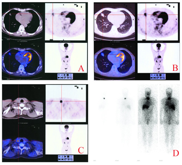
A 56-year-old man with coughing three months. FDG PET/CT was performed, and resection specimens of lung and thyroid were detected by hematoxylin eosin staining (HE) and IHC. PET/CT: lung tumor SUVmax: 3.69, delay: 5.17; and thyroid tumor SUVmax 19.97. HE reveal papillary adenocarcinoma, but histological differentiation of primary pulmonary adenocarcinoma from metastatic adenocarcinoma is sometimes difficult because of their phenotypic similarities. So IHC was performed, the IHC of lung tumor: cytokeratin 20 (CK20)⁻, thyroglobulin(Tg)⁻, cytokeratin7(CK7)+, thyroid transcription factor-1 (TTF-1)+; thyroid tumor: CK7+, TTF-1+, thyroglobulin+, CK20⁻. Therefore, the final diagnosis was double primary adenocarcinomas of thyroid and lung.
FDG PET/CT has preliminary diagnostic capacity of multiple primary tumors; the final diagnosis should be adopted for specimens after tumor-specific markers IHC to obtain. Consequently, effective therapeutic approaches can be designed and conducted.
甲状腺和肺双原发乳头状腺癌的准确诊断更为罕见,据我们所知,尚无文献报道通过 [¹⁸F]-2-氟-2-脱氧-D-葡萄糖正电子发射断层扫描/X 射线计算机断层扫描(FDG PET/CT)结合手术标本免疫组织化学(IHC)对其进行诊断。我们报告了一例 FDG PET/CT 示甲状腺和肺异常的患者,这种不典型表现若无手术标本 IHC 可能导致误诊。
一名 56 岁男性,咳嗽 3 个月。行 FDG PET/CT 检查,对肺和甲状腺的切除标本进行了苏木精-伊红(HE)染色和 IHC 检测。PET/CT:肺肿瘤 SUVmax:3.69,延迟:5.17;甲状腺肿瘤 SUVmax:19.97。HE 显示为乳头状腺癌,但由于其表型相似,原发性肺腺癌与转移性腺癌的组织学分化有时较为困难。因此进行了 IHC 检测,肺肿瘤的 IHC:细胞角蛋白 20(CK20)⁻,甲状腺球蛋白(Tg)⁻,细胞角蛋白 7(CK7)⁺,甲状腺转录因子-1(TTF-1)⁺;甲状腺肿瘤:CK7⁺,TTF-1⁺,甲状腺球蛋白⁺,CK20⁻。因此,最终诊断为甲状腺和肺双原发腺癌。
FDG PET/CT 对多原发肿瘤具有初步诊断能力;最终诊断应采用肿瘤特异性标志物 IHC 后的标本获得。从而可以制定和实施有效的治疗方法。