Department of Molecular Pharmacology, Graduate School of Pharmaceutical Sciences, Kyoto University, Kyoto, Japan.
PLoS One. 2011;6(9):e24865. doi: 10.1371/journal.pone.0024865. Epub 2011 Sep 30.
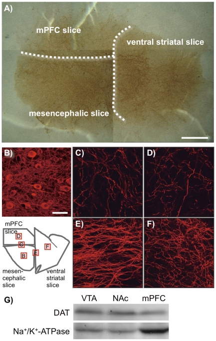
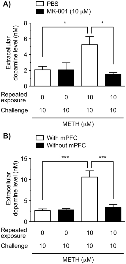
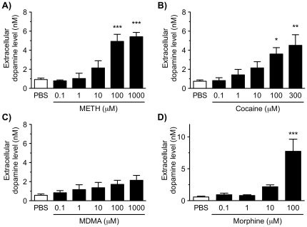
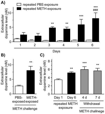
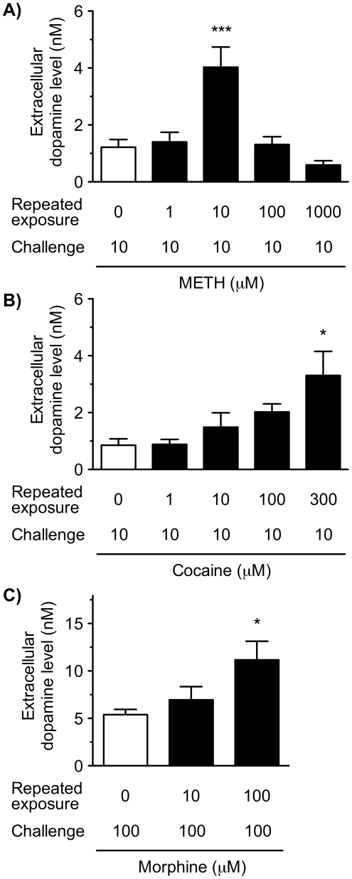
Repeated intermittent exposure to psychostimulants and morphine leads to progressive augmentation of its locomotor activating effects in rodents. Accumulating evidence suggests the critical involvement of the mesocorticolimbic dopaminergic neurons, which project from the ventral tegmental area to the nucleus accumbens and the medial prefrontal cortex, in the behavioral sensitization. Here, we examined the acute and chronic effects of psychostimulants and morphine on dopamine release in a reconstructed mesocorticolimbic system comprised of a rat triple organotypic slice co-culture of the ventral tegmental area, nucleus accumbens and medial prefrontal cortex regions. Tyrosine hydroxylase-positive cell bodies were localized in the ventral tegmental area, and their neurites projected to the nucleus accumbens and medial prefrontal cortex regions. Acute treatment with methamphetamine (0.1-1000 µM), cocaine (0.1-300 µM) or morphine (0.1-100 µM) for 30 min increased extracellular dopamine levels in a concentration-dependent manner, while 3,4-methylenedioxyamphetamine (0.1-1000 µM) had little effect. Following repeated exposure to methamphetamine (10 µM) for 30 min every day for 6 days, the dopamine release gradually increased during the 30-min treatment. The augmentation of dopamine release was maintained even after the withdrawal of methamphetamine for 7 days. Similar augmentation was observed by repeated exposure to cocaine (1-300 µM) or morphine (10 and 100 µM). Furthermore, methamphetamine-induced augmentation of dopamine release was prevented by an NMDA receptor antagonist, MK-801 (10 µM), and was not observed in double slice co-cultures that excluded the medial prefrontal cortex slice. These results suggest that repeated psychostimulant- or morphine-induced augmentation of dopamine release, i.e. dopaminergic sensitization, was reproduced in a rat triple organotypic slice co-cultures. In addition, the slice co-culture system revealed that the NMDA receptors and the medial prefrontal cortex play an essential role in the dopaminergic sensitization. This in vitro sensitization model provides a unique approach for studying mechanisms underlying behavioral sensitization to drugs of abuse.
反复间歇性暴露于精神兴奋剂和吗啡会导致其在啮齿动物中的运动激活作用逐渐增强。越来越多的证据表明,从中脑边缘多巴胺能神经元的关键参与,其从腹侧被盖区投射到伏隔核和内侧前额叶皮层,在行为敏感化。在这里,我们检查了精神兴奋剂和吗啡对包含腹侧被盖区、伏隔核和内侧前额叶皮层区域的大鼠三重器官型切片共培养物的中脑边缘多巴胺释放的急性和慢性影响。酪氨酸羟化酶阳性细胞体位于腹侧被盖区,其神经突投射到伏隔核和内侧前额叶皮层区域。急性治疗用甲基苯丙胺(0.1-1000μM),可卡因(0.1-300μM)或吗啡(0.1-100μM)30 分钟增加细胞外多巴胺水平呈浓度依赖性,而 3,4-亚甲二氧基苯丙胺(0.1-1000μM)几乎没有效果。在重复暴露于甲基苯丙胺(10μM)30 分钟,每天 6 天后,多巴胺释放逐渐增加在 30 分钟的治疗过程中。即使在停止甲基苯丙胺 7 天后,多巴胺释放的增加仍保持不变。重复暴露于可卡因(1-300μM)或吗啡(10 和 100μM)也观察到类似的增强。此外,NMDA 受体拮抗剂 MK-801(10μM)阻止了甲基苯丙胺诱导的多巴胺释放增加,并且在排除了内侧前额叶皮层切片的双切片共培养物中未观察到。这些结果表明,重复的精神兴奋剂或吗啡诱导的多巴胺释放增加,即多巴胺能敏感化,在大鼠三重器官型切片共培养物中得到重现。此外,切片共培养系统表明 NMDA 受体和内侧前额叶皮层在多巴胺能敏感化中起重要作用。这种体外敏感化模型为研究滥用药物行为敏感化的机制提供了一种独特的方法。