Keenan Research Centre of the Li Ka Shing Knowledge Institute, St. Michael's Hospital, Toronto, Ontario, Canada.
PLoS One. 2011;6(10):e24695. doi: 10.1371/journal.pone.0024695. Epub 2011 Oct 3.
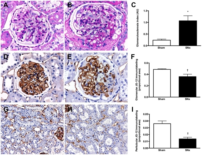
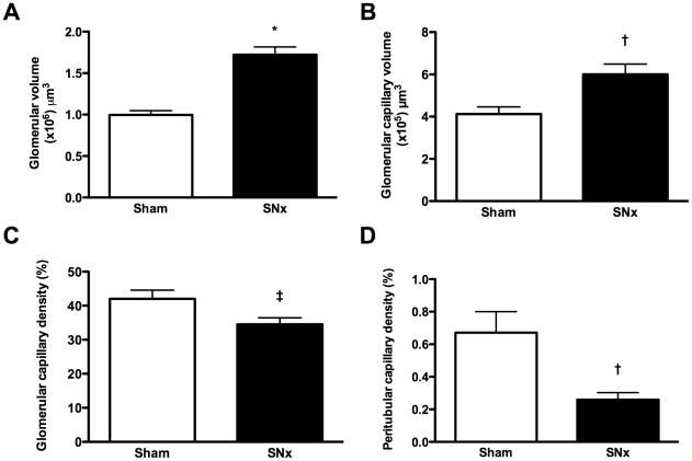
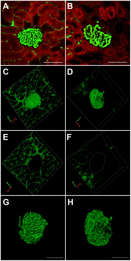
Rarefaction of the renal microvasculature correlates with declining kidney function. However, current technologies commonly used for its evaluation are limited by their reliance on endothelial cell antigen expression and assessment in two dimensions. We set out to establish a widely applicable and unbiased optical sectioning method to enable three dimensional imaging and reconstruction of the renal microvessels based on their luminal filling. The kidneys of subtotally nephrectomized (SNx) rats and their sham-operated counterparts were subjected to either routine two-dimensional immunohistochemistry or the novel technique of fluorescent microangiography (FMA). The latter was achieved by perfusion of the kidney with an agarose suspension of fluorescent polystyrene microspheres followed by optical sectioning of 200 µm thick cross-sections using a confocal microscope. The fluorescent microangiography method enabled the three-dimensional reconstruction of virtual microvascular casts and confirmed a reduction in both glomerular and peritubular capillary density in the kidneys of SNx rats, despite an overall increase in glomerular volume. FMA is an uncomplicated technique for evaluating the renal microvasculature that circumvents many of the limitations imposed by conventional analysis of two-dimensional tissue sections.
肾微血管稀疏与肾功能下降相关。然而,目前常用于评估肾微血管稀疏的技术受到其对内皮细胞抗原表达的依赖以及二维评估的限制。我们旨在建立一种广泛适用且无偏倚的光学切片方法,以便能够基于管腔充盈对肾微血管进行三维成像和重建。对部分肾切除 (SNx) 大鼠及其假手术对照大鼠的肾脏进行常规二维免疫组织化学或新型荧光微血管造影 (FMA) 检测。后者通过用琼脂糖悬浮的荧光聚苯乙烯微球对肾脏进行灌注,然后使用共聚焦显微镜对 200 µm 厚的横截面进行光学切片来实现。荧光微血管造影方法能够对虚拟微血管铸型进行三维重建,并证实尽管肾小球总体积增加,但 SNx 大鼠的肾小球和肾小管周围毛细血管密度均降低。FMA 是一种评估肾微血管的简单技术,它规避了传统二维组织切片分析所带来的许多限制。