• 文献检索
  • 文档翻译
  • 深度研究
  • 学术资讯
  • Suppr Zotero 插件Zotero 插件
  • 邀请有礼
  • 套餐&价格
  • 历史记录
应用&插件
Suppr Zotero 插件Zotero 插件浏览器插件Mac 客户端Windows 客户端微信小程序
定价
高级版会员购买积分包购买API积分包
服务
文献检索文档翻译深度研究API 文档MCP 服务
关于我们
关于 Suppr公司介绍联系我们用户协议隐私条款
关注我们

Suppr 超能文献

核心技术专利:CN118964589B侵权必究
粤ICP备2023148730 号-1Suppr @ 2026

文献检索

告别复杂PubMed语法,用中文像聊天一样搜索,搜遍4000万医学文献。AI智能推荐,让科研检索更轻松。

立即免费搜索

文件翻译

保留排版,准确专业,支持PDF/Word/PPT等文件格式,支持 12+语言互译。

免费翻译文档

深度研究

AI帮你快速写综述,25分钟生成高质量综述,智能提取关键信息,辅助科研写作。

立即免费体验

输送技术在确定 Enterprise 自膨式颅内支架的血管壁贴壁方面起着重要作用。

Delivery technique plays an important role in determining vessel wall apposition of the Enterprise self-expanding intracranial stent.

机构信息

Cerebrovascular and Endovascular Division, Department of Neurosurgery, Tufts Medical Center, Boston, Massachusetts 02111, USA.

出版信息

J Neurointerv Surg. 2011 Dec 1;3(4):340-3. doi: 10.1136/jnis.2010.004499. Epub 2011 Mar 9.

DOI:10.1136/jnis.2010.004499
PMID:21990459
原文链接:https://pmc.ncbi.nlm.nih.gov/articles/PMC3212648/
Abstract

BACKGROUND

The Enterprise (EN) vascular reconstruction device is a self-expanding nitinol stent used as adjunctive support in wide-necked aneurysm coiling. We sought to evaluate the effect of deployment technique on how well the EN stent conforms to the vessel wall around a curve.

METHODS

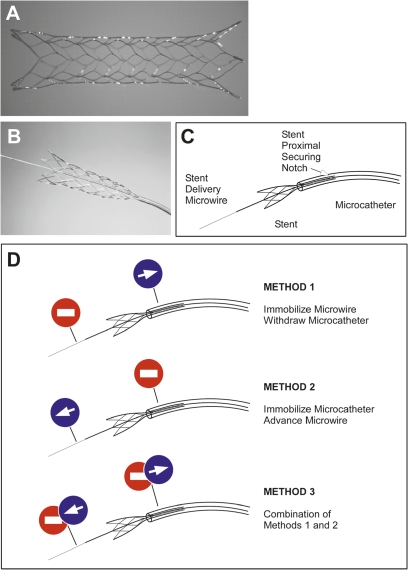
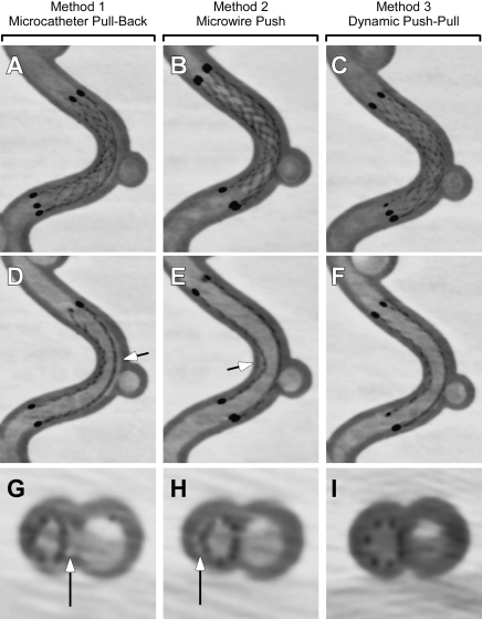
A flow model consisting of a 3.5 mm diameter silicone tube forming a 7 mm radius curve was visualized using high-resolution flat-panel CT (FPCT; DynaCT). EN stents (4.5 mm × 22 mm) were deployed using three methods: (1) microcatheter pull-back, (2) delivery microwire push and (3) a combination of both methods so as to keep the microcatheter tip centered within the lumen during deployment. FPCT images were visualized using multiplanar reconstruction for evidence of incomplete stent apposition (ISA).

RESULTS

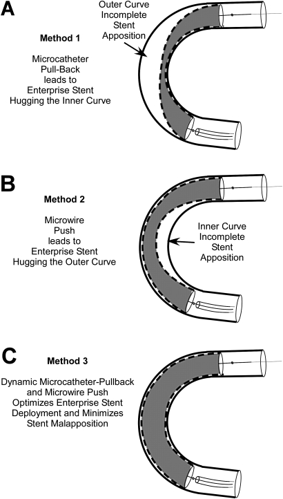
FPCT revealed a critical role for deployment method in stent-wall apposition as noted by the development of a crescent-shaped gap between the stent and the wall. Specifically, the manufacturer-recommended microcatheter pull-back unsheathing technique (method 1) resulted in outer curve ISA, while the microwire push technique (method 2) led to inner curve ISA. Using method 3 in a dynamic push-pull manner minimized both inner and outer curve ISA.

CONCLUSION

The deployment method used to deliver the EN vascular reconstruction device plays a critical role in determining how well its struts appose the vessel wall in vitro. This characteristic must be taken into account when deploying this flexible low-profile stent to avoid ISA in even mildly tortuous anatomy given the possible link between stent malapposition and thromboembolic complications.

摘要

背景

Enterprise(EN)血管重建装置是一种自膨式镍钛诺支架,用作宽颈动脉瘤线圈的辅助支撑。我们旨在评估部署技术对 EN 支架在曲线上与血管壁贴合程度的影响。

方法

使用高分辨率平板 CT(FPCT;DynaCT)可视化由 3.5 毫米直径硅树脂管形成 7 毫米半径曲线的流模型。使用三种方法部署 EN 支架(4.5 毫米×22 毫米):(1)微导管回缩,(2)输送微丝推送,(3)结合这两种方法,以便在部署过程中使微导管尖端保持在管腔中心。使用多平面重建可视化 FPCT 图像,以证明支架壁贴合不完全(ISA)。

结果

FPCT 揭示了部署方法在支架壁贴合中的关键作用,这表现在支架和壁之间形成新月形间隙。具体来说,制造商推荐的微导管回缩解鞘技术(方法 1)导致外曲线 ISA,而微丝推送技术(方法 2)导致内曲线 ISA。以动态推-拉方式使用方法 3可最大限度地减少内曲线和外曲线 ISA。

结论

用于输送 EN 血管重建装置的部署方法在确定其支柱在体外与血管壁贴合程度方面起着关键作用。在部署这种灵活的低轮廓支架时,必须考虑到这一特性,以避免在轻度扭曲的解剖结构中发生 ISA,因为支架贴壁不良与血栓栓塞并发症之间可能存在关联。

https://cdn.ncbi.nlm.nih.gov/pmc/blobs/a240/3212648/5010e6011447/neurintsurg4499fig3.jpg
https://cdn.ncbi.nlm.nih.gov/pmc/blobs/a240/3212648/e818da785ffa/neurintsurg4499fig1.jpg
https://cdn.ncbi.nlm.nih.gov/pmc/blobs/a240/3212648/868b1a11fd8e/neurintsurg4499fig2.jpg
https://cdn.ncbi.nlm.nih.gov/pmc/blobs/a240/3212648/5010e6011447/neurintsurg4499fig3.jpg
https://cdn.ncbi.nlm.nih.gov/pmc/blobs/a240/3212648/e818da785ffa/neurintsurg4499fig1.jpg
https://cdn.ncbi.nlm.nih.gov/pmc/blobs/a240/3212648/868b1a11fd8e/neurintsurg4499fig2.jpg
https://cdn.ncbi.nlm.nih.gov/pmc/blobs/a240/3212648/5010e6011447/neurintsurg4499fig3.jpg

相似文献

1
Delivery technique plays an important role in determining vessel wall apposition of the Enterprise self-expanding intracranial stent.输送技术在确定 Enterprise 自膨式颅内支架的血管壁贴壁方面起着重要作用。
J Neurointerv Surg. 2011 Dec 1;3(4):340-3. doi: 10.1136/jnis.2010.004499. Epub 2011 Mar 9.
2
Parent vessel size and curvature strongly influence risk of incomplete stent apposition in enterprise intracranial aneurysm stent coiling.载瘤动脉的大小和弯曲度强烈影响 Enterprise 颅内动脉瘤支架圈闭术中支架贴壁不完全的风险。
AJNR Am J Neuroradiol. 2011 Oct;32(9):1714-20. doi: 10.3174/ajnr.A2584. Epub 2011 Aug 4.
3
Deployment technique that takes advantage of the characteristics of Enterprise VRD2: an in vitro study.利用Enterprise VRD2特性的部署技术:一项体外研究。
J Neurointerv Surg. 2017 Oct;9(10):969-973. doi: 10.1136/neurintsurg-2016-012400. Epub 2016 Aug 31.
4
In vitro experiments of vessel wall apposition between the Enterprise and Enterprise 2 stents for treatment of cerebral aneurysms.用于治疗脑动脉瘤的Enterprise和Enterprise 2支架之间血管壁贴合的体外实验。
Acta Neurochir (Wien). 2016 Feb;158(2):241-5. doi: 10.1007/s00701-015-2666-3. Epub 2015 Dec 19.
5
Double-barrel entanglement of intracranial Enterprise stents resulting from undetected incomplete stent apposition.由于未检测到不完全的支架贴壁,导致颅内 Enterprise 支架的双筒状缠绕。
J Neurointerv Surg. 2012 Nov;4(6):e38. doi: 10.1136/neurintsurg-2011-010192. Epub 2012 Jan 9.
6
Active push deployment technique improves stent/vessel-wall interaction in endovascular treatment of acute stroke with stent retrievers.主动推送部署技术改善了使用取栓支架进行急性卒中血管内治疗时支架与血管壁的相互作用。
J Neurointerv Surg. 2017 Mar;9(3):253-256. doi: 10.1136/neurintsurg-2016-012322. Epub 2016 Mar 14.
7
Incomplete stent apposition of low-profile visualized intraluminal support stents in the treatment of cerebral aneurysms.低轮廓可视化腔内支撑支架在治疗脑动脉瘤中支架贴壁不完整的情况。
J Neurointerv Surg. 2020 Jun;12(6):591-597. doi: 10.1136/neurintsurg-2019-015505. Epub 2019 Dec 9.
8
DynaCT imaging for intraprocedural evaluation of flow-diverting stent apposition during endovascular treatment of intracranial aneurysms.在颅内动脉瘤血管内治疗期间,使用动态容积CT成像进行血流导向支架贴壁的术中评估。
J Clin Neurosci. 2014 Nov;21(11):1981-3. doi: 10.1016/j.jocn.2014.04.003. Epub 2014 May 20.
9
Crescent sign on magnetic resonance angiography revealing incomplete stent apposition: correlation with diffusion-weighted changes in stent-mediated coil embolization of aneurysms.磁共振血管造影上的新月征提示支架贴壁不完全:与支架介导的动脉瘤线圈栓塞术所致的弥散加权变化的相关性。
J Neurosurg. 2011 Sep;115(3):624-32. doi: 10.3171/2011.4.JNS102050. Epub 2011 May 27.
10
The Enterprise2 Stent for Endovascular Treatment of Intracranial Aneurysms: Short-Term Results From a Single Center Experience.用于颅内动脉瘤血管内治疗的Enterprise2支架:单中心经验的短期结果
Front Neurol. 2020 Dec 3;11:589689. doi: 10.3389/fneur.2020.589689. eCollection 2020.

引用本文的文献

1
Flow-diverter stents combined with flow-T stenting-assisted coiling for the treatment of a large basilar apex aneurysm: a case report with a 9-month follow-up.血流导向支架联合血流T型支架辅助弹簧圈栓塞治疗大型基底动脉尖动脉瘤:一例9个月随访的病例报告
Front Neurol. 2024 Jan 19;14:1247549. doi: 10.3389/fneur.2023.1247549. eCollection 2023.
2
Evaluation of Stent Apposition in the LVIS Blue Stent-Assisted Coiling of Distal Internal Carotid Artery Aneurysms : Correlation with Clinical and Angiographic Outcomes.远端颈内动脉动脉瘤LVIS蓝色支架辅助弹簧圈栓塞中支架贴壁情况的评估:与临床和血管造影结果的相关性
J Korean Neurosurg Soc. 2022 Nov;65(6):801-815. doi: 10.3340/jkns.2022.0021. Epub 2022 Jun 22.
3

本文引用的文献

1
Strut apposition after coronary stent implantation visualised with optical coherence tomography.光学相干断层成像术观察到冠状动脉支架植入术后的支架贴壁不良。
EuroIntervention. 2010 May;6(1):86-93.
2
Incomplete stent apposition after coronary stent implantation: myth or reality?冠状动脉支架植入术后支架贴壁不全:是误解还是确有其事?
J Interv Cardiol. 2009 Aug;22(4):341-9. doi: 10.1111/j.1540-8183.2009.00485.x.
3
Spontaneous delayed proximal migration of enterprise stent after staged treatment of wide-necked basilar aneurysm: technical case report.
"Virtual stent" - clinical evaluation and user experience of on-the-fly stent simulation in treatment of cerebral aneurysms.
“虚拟支架”——血流导向装置在颅内动脉瘤治疗中的即时支架模拟的临床评估和用户体验。
Interv Neuroradiol. 2022 Oct;28(5):581-587. doi: 10.1177/15910199211053131. Epub 2021 Oct 29.
4
Combination of high-resolution cone beam computed tomography and metal artefact reduction software: a new image fusion technique for evaluating intracranial stent apposition after aneurysm treatment.高分辨率锥形束计算机断层扫描与金属伪影减少软件的结合:一种评估动脉瘤治疗后颅内支架贴壁情况的新型图像融合技术。
BMJ Case Rep. 2019 Sep 17;12(9):e230687. doi: 10.1136/bcr-2019-230687.
5
Accuracy of Length of Virtual Stents in Treatment of Intracranial Wide-Necked Aneurysms.颅内宽颈动脉瘤治疗中虚拟支架长度的准确性。
Cardiovasc Intervent Radiol. 2019 Aug;42(8):1168-1174. doi: 10.1007/s00270-019-02230-9. Epub 2019 May 10.
6
Endovascular Coiling for a Wide-neck Bifurcated Aneurysm with Anterograde Horizontal Stenting via Microcatheter Looping: A Technical Case Report.经微导管成袢顺行水平支架置入术治疗宽颈分叉动脉瘤的血管内栓塞:技术病例报告
J Cerebrovasc Endovasc Neurosurg. 2018 Sep;20(3):181-186. doi: 10.7461/jcen.2018.20.3.181. Epub 2018 Sep 30.
7
The safety and effectiveness of the LVIS stent system for the treatment of wide-necked cerebral aneurysms: final results of the pivotal US LVIS trial.LVIS 支架系统治疗宽颈颅内动脉瘤的安全性和有效性:美国 LVIS 关键性试验的最终结果。
J Neurointerv Surg. 2019 Apr;11(4):357-361. doi: 10.1136/neurintsurg-2018-014309. Epub 2018 Oct 8.
8
Herniated Proximal Marker of the Stent into the Meningohypophyseal Trunk after Stent-Assisted Coil Embolization for Intracranial Aneurysms.颅内动脉瘤支架辅助弹簧圈栓塞术后支架进入脑膜垂体干的近端疝出标记物。
Case Rep Neurol. 2018 Aug 29;10(2):213-216. doi: 10.1159/000492110. eCollection 2018 May-Aug.
9
A comparison between the new Low-profile Visualized Intraluminal Support (LVIS Blue) stent and the Flow Redirection Endoluminal Device (FRED) in bench-top and cadaver studies.新型低轮廓可视化腔内支撑(LVIS Blue)支架与血流导向装置(FRED)在台架和尸体研究中的比较。
J Neurointerv Surg. 2018 Mar;10(3):274-278. doi: 10.1136/neurintsurg-2017-013074. Epub 2017 Sep 22.
10
Bench-top Comparison of Physical Properties of 4 Commercially-Available Self-Expanding Intracranial Stents.4种市售自膨式颅内支架物理特性的台式比较
Neurointervention. 2017 Mar;12(1):31-39. doi: 10.5469/neuroint.2017.12.1.31. Epub 2017 Mar 6.
宽颈基底动脉瘤分期治疗后Enterprise支架自发延迟性近端移位:技术病例报告
Neurosurgery. 2009 May;64(5):E1012; discussion E1012. doi: 10.1227/01.NEU.0000343745.18753.D6.
4
Treatment of intracranial aneurysms with the Enterprise stent: a multicenter registry.使用Enterprise支架治疗颅内动脉瘤:一项多中心注册研究。
J Neurosurg. 2009 Jan;110(1):35-9. doi: 10.3171/2008.7.JNS08322.
5
Delayed migration of a self-expanding intracranial microstent.自膨式颅内微支架延迟移位
AJNR Am J Neuroradiol. 2008 Nov;29(10):1959-60. doi: 10.3174/ajnr.A1224. Epub 2008 Aug 21.
6
Stent conformity in curved vascular models with simulated aneurysm necks using flat-panel CT: an in vitro study.使用平板CT在具有模拟动脉瘤颈部的弯曲血管模型中进行支架贴合度的体外研究。
AJNR Am J Neuroradiol. 2007 May;28(5):823-9.
7
Increased cell opening and prolapse of struts of a neuroform stent in curved vasculature: value of angiographic computed tomography: technical case report.
Neurosurgery. 2006 Apr;58(4 Suppl 2):ONS-E380; discussion ONS-E380. doi: 10.1227/01.NEU.0000205287.06739.E1.
8
Initial clinical experience with a new self-expanding nitinol stent for the treatment of intracranial cerebral aneurysms: the Cordis Enterprise stent.一种用于治疗颅内脑动脉瘤的新型自膨式镍钛合金支架——科迪斯企业支架的初步临床经验
AJNR Am J Neuroradiol. 2005 Aug;26(7):1751-6.
9
The Neuroform stent, the first microcatheter-delivered stent for use in the intracranial circulation.Neuroform支架是首个通过微导管输送用于颅内循环的支架。
Neurosurgery. 2004 Jan;54(1):2-5. doi: 10.1227/01.neu.0000099370.05758.4d.