Department of Morphological Brain Science, Graduate School of Medicine, Kyoto University Kyoto, Japan.
Front Neural Circuits. 2011 Sep 29;5:12. doi: 10.3389/fncir.2011.00012. eCollection 2011.
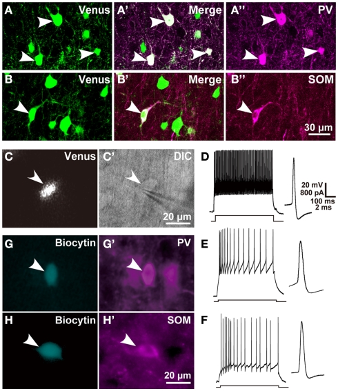
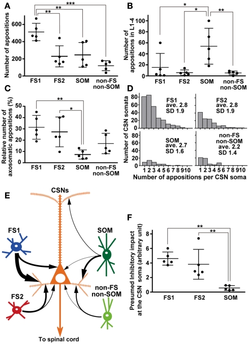
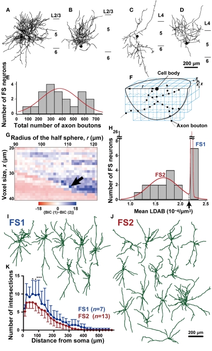
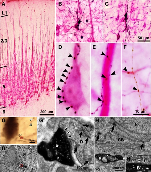
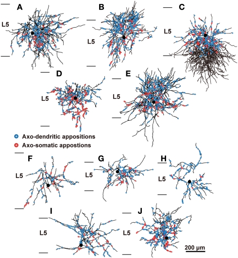
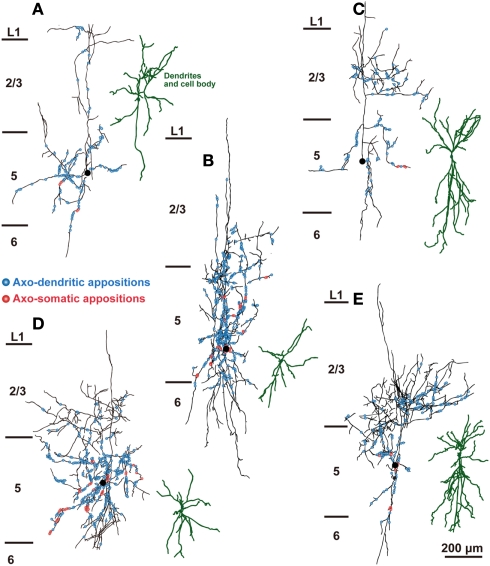
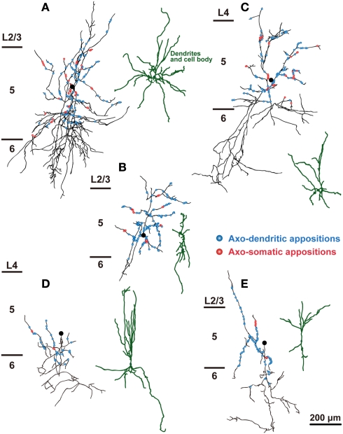
In the local circuit of the cerebral cortex, GABAergic inhibitory interneurons are considered to work in collaboration with excitatory neurons. Although many interneuron subgroups have been described in the cortex, local inhibitory connections of each interneuron subgroup are only partially understood with respect to the functional neuron groups that receive these inhibitory connections. In the present study, we morphologically examined local inhibitory inputs to corticospinal neurons (CSNs) in motor areas using transgenic rats in which GABAergic neurons expressed fluorescent protein Venus. By analysis of biocytin-filled axons obtained with whole-cell recording/staining in cortical slices, we classified fast-spiking (FS) neurons in layer (L) 5 into two types, FS1 and FS2, by their high and low densities of axonal arborization, respectively. We then investigated the connections of FS1, FS2, somatostatin (SOM)-immunopositive, and other (non-FS/non-SOM) interneurons to CSNs that were retrogradely labeled in motor areas. When close appositions between the axon boutons of the intracellularly labeled interneurons and the somata/dendrites of the retrogradely labeled CSNs were examined electron-microscopically, 74% of these appositions made symmetric synaptic contacts. The axon boutons of single FS1 neurons were two- to fourfold more frequent in appositions to the somata/dendrites of CSNs than those of FS2, SOM, and non-FS/non-SOM neurons. Axosomatic appositions were most frequently formed with axon boutons of FS1 and FS2 neurons (approximately 30%) and least frequently formed with those of SOM neurons (7%). In contrast, SOM neurons most extensively sent axon boutons to the apical dendrites of CSNs. These results might suggest that motor outputs are controlled differentially by the subgroups of L5 GABAergic interneurons in cortical motor areas.
在大脑皮层的局部回路中,GABA 能抑制性中间神经元被认为与兴奋性神经元协同工作。尽管皮层中已经描述了许多中间神经元亚群,但对于每个中间神经元亚群的局部抑制性连接,仅部分了解其接收这些抑制性连接的功能神经元群。在本研究中,我们使用在皮层中表达荧光蛋白 Venus 的转基因大鼠,对运动区的皮质脊髓神经元(CSN)的局部抑制性输入进行了形态学检查。通过对皮层切片中全细胞记录/染色获得的生物胞素填充轴突进行分析,我们根据轴突分支的高密度和低密度,将 L5 中的快速放电(FS)神经元分别分为 FS1 和 FS2 两种类型。然后,我们研究了 FS1、FS2、生长抑素(SOM)免疫阳性和其他(非 FS/非 SOM)中间神经元与在运动区中逆行标记的 CSN 的连接。当观察到用细胞内标记的中间神经元的轴突末梢与逆行标记的 CSN 的胞体/树突之间的紧密贴合时,74%的这些贴合形成了对称的突触接触。单个 FS1 神经元的轴突末梢与 CSN 的胞体/树突的贴合次数是 FS2、SOM 和非 FS/非 SOM 神经元的两到四倍。轴突末梢与 FS1 和 FS2 神经元(约 30%)形成的轴突末梢与 FS1 和 FS2 神经元的贴合最为频繁,与 SOM 神经元的贴合最少(7%)。相比之下,SOM 神经元最广泛地将轴突末梢发送到 CSN 的树突。这些结果可能表明,运动输出是由皮层运动区的 L5 GABA 能中间神经元亚群差异控制的。