Edwards Malia M, McLeod D Scott, Grebe Rhonda, Heng Céline, Lefebvre Olivier, Lutty Gerard A
The Wilmer Eye Institute, Baltimore, MD 21287, USA.
BMC Dev Biol. 2011 Oct 14;11:60. doi: 10.1186/1471-213X-11-60.
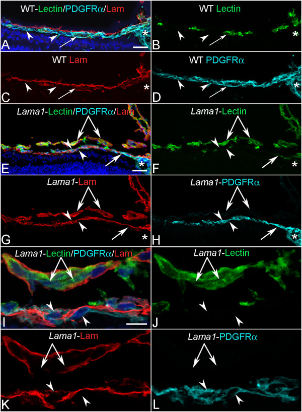
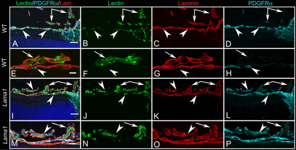
Valuable insights into the complex process of retinal vascular development can be gained using models with abnormal retinal vasculature. Two such models are the recently described mouse lines with mutations in Lama1, an important component of the retinal internal limiting membrane (ILM). These mutants have a persistence of the fetal vasculature of vitreous (FVV) but lack a primary retinal vascular plexus. The present study provides a detailed analysis of astrocyte and vascular development in these Lama1 mutants.
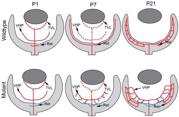
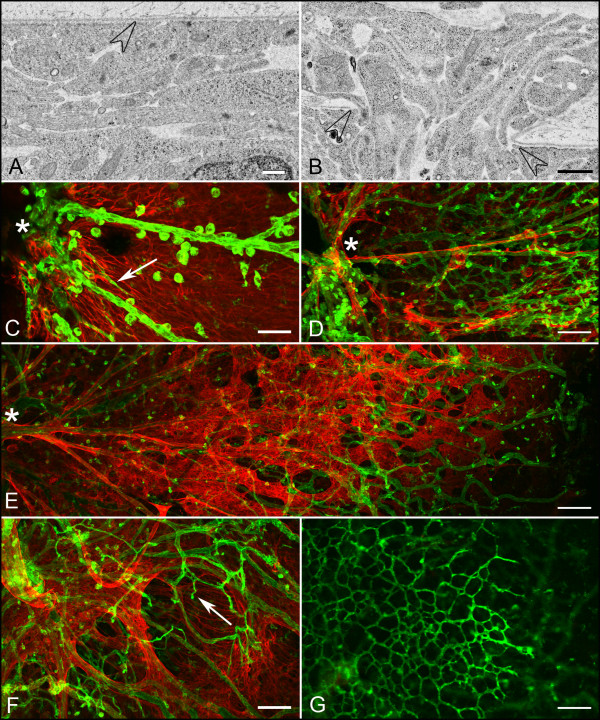
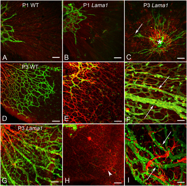
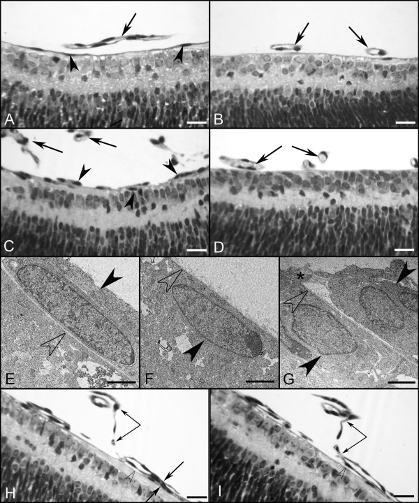
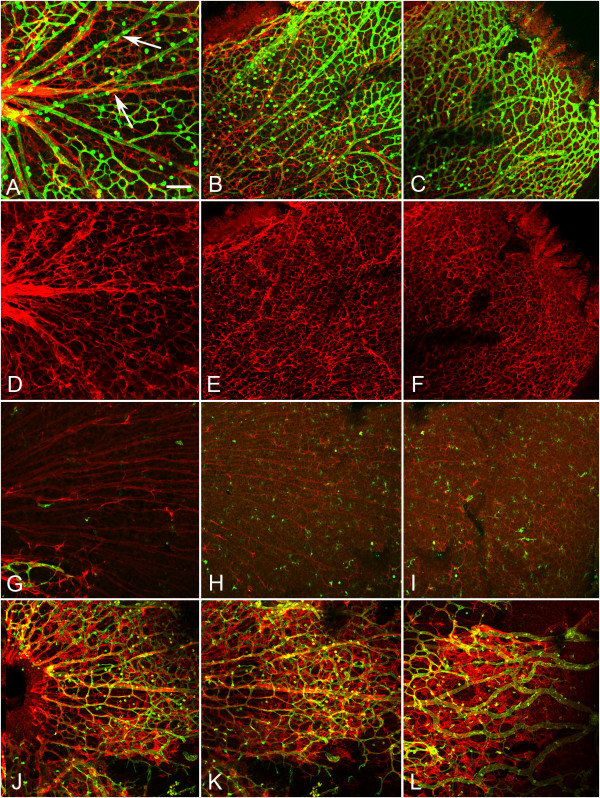
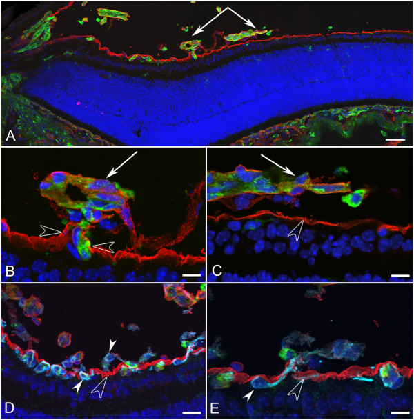
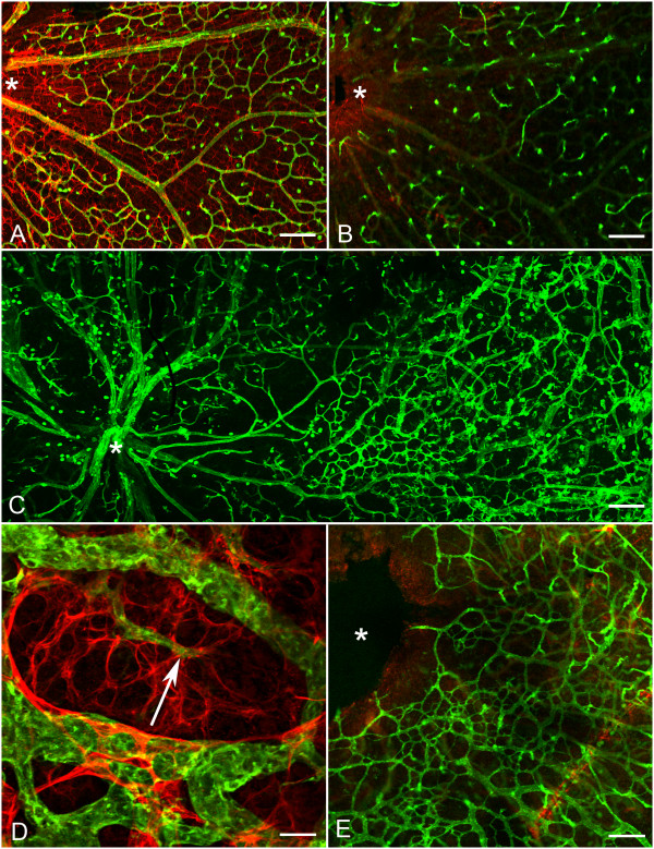
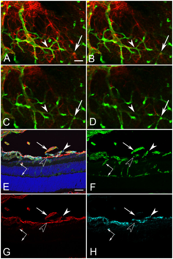
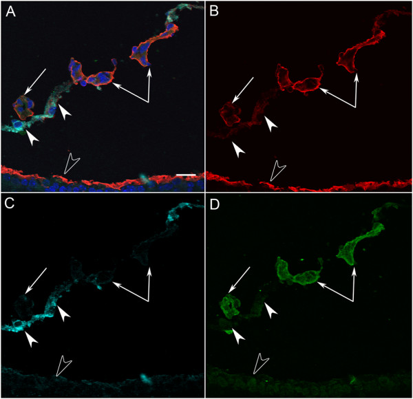
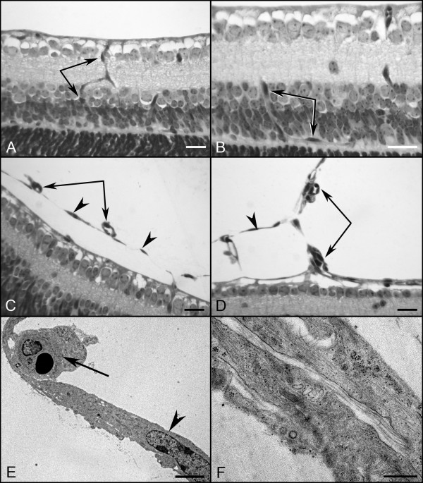
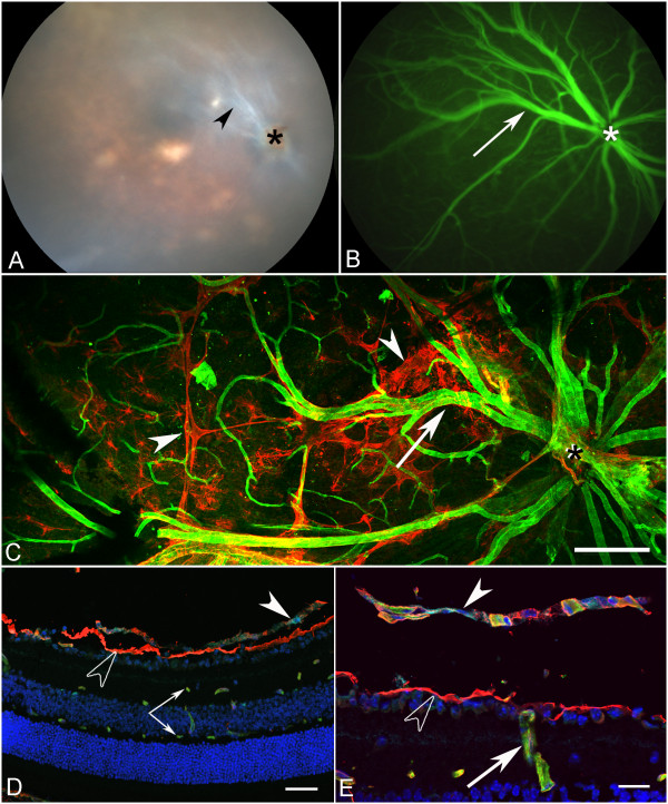
Although astrocytes and blood vessels initially migrate into Lama1 mutant retinas, both traverse the peripapillary ILM into the vitreous by P3. Once in the vitreous, blood vessels anastomose with vessels of the vasa hyaloidea propria, part of the FVV, and eventually re-enter the retina where they dive to form the inner and outer retinal capillary networks. Astrocytes continue proliferating within the vitreous to form a dense mesh that resembles epiretinal membranes associated with persistent fetal vasculature and proliferative vitreoretinopathy.
Lama1 and a fully intact ILM are required for normal retinal vascular development. Mutations in Lama1 allow developing retinal vessels to enter the vitreous where they anastomose with vessels of the hyaloid system which persist and expand. Together, these vessels branch into the retina to form fairly normal inner retinal vascular capillary plexi. The Lama1 mutants described in this report are potential models for studying the human conditions persistent fetal vasculature and proliferative vitreoretinopathy.
利用视网膜血管异常的模型,可深入了解视网膜血管发育的复杂过程。其中两个这样的模型是最近描述的Lama1发生突变的小鼠品系,Lama1是视网膜内界膜(ILM)的重要组成部分。这些突变体存在玻璃体胎儿血管(FVV)持续存在的情况,但缺乏原发性视网膜血管丛。本研究对这些Lama1突变体中的星形胶质细胞和血管发育进行了详细分析。
虽然星形胶质细胞和血管最初迁移到Lama1突变体视网膜中,但两者在出生后第3天(P3)均穿过视乳头周围的内界膜进入玻璃体。一旦进入玻璃体,血管就会与固有玻璃体血管(FVV的一部分)的血管吻合,最终重新进入视网膜,在那里它们向下延伸形成视网膜内、外毛细血管网络。星形胶质细胞在玻璃体内持续增殖,形成一个致密的网状结构,类似于与持续性胎儿血管和增殖性玻璃体视网膜病变相关的视网膜前膜。
正常的视网膜血管发育需要Lama1和完整的内界膜。Lama1的突变使发育中的视网膜血管进入玻璃体,在那里它们与持续存在并扩张的玻璃体系统血管吻合。这些血管一起分支进入视网膜,形成相当正常的视网膜内血管毛细血管丛。本报告中描述的Lama1突变体是研究人类持续性胎儿血管和增殖性玻璃体视网膜病变的潜在模型。