• 文献检索
  • 文档翻译
  • 深度研究
  • 学术资讯
  • Suppr Zotero 插件Zotero 插件
  • 邀请有礼
  • 套餐&价格
  • 历史记录
应用&插件
Suppr Zotero 插件Zotero 插件浏览器插件Mac 客户端Windows 客户端微信小程序
定价
高级版会员购买积分包购买API积分包
服务
文献检索文档翻译深度研究API 文档MCP 服务
关于我们
关于 Suppr公司介绍联系我们用户协议隐私条款
关注我们

Suppr 超能文献

核心技术专利:CN118964589B侵权必究
粤ICP备2023148730 号-1Suppr @ 2026

文献检索

告别复杂PubMed语法,用中文像聊天一样搜索,搜遍4000万医学文献。AI智能推荐,让科研检索更轻松。

立即免费搜索

文件翻译

保留排版,准确专业,支持PDF/Word/PPT等文件格式,支持 12+语言互译。

免费翻译文档

深度研究

AI帮你快速写综述,25分钟生成高质量综述,智能提取关键信息,辅助科研写作。

立即免费体验

儿科肿瘤影像学中的陷阱。

Pitfalls in paediatric oncology imaging.

机构信息

Radiology Department, Great Ormond Street Hospital for Children, London, UK.

出版信息

Cancer Imaging. 2011 Sep 24;11(1):144-54. doi: 10.1102/1470-7330.2011.0021.

DOI:10.1102/1470-7330.2011.0021
PMID:22004902
原文链接:https://pmc.ncbi.nlm.nih.gov/articles/PMC3205763/
Abstract

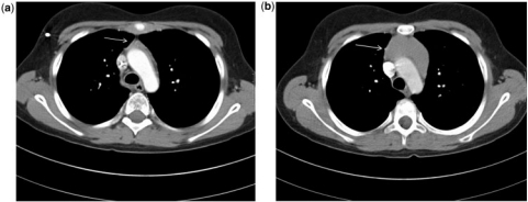
Imaging a new mass lesion in a child requires careful consideration of a variety of issues. The age of the child is an important factor in determining the appropriate test to start with and the age also helps provide an appropriate differential diagnosis, which can then be used to guide further imaging. The long-term outcome for most children with cancer is very good, with over 70% achieving 5-year survival and presumed cure. Consequently their imaging requirements should be regarded as equal to all other children. Minimizing exposure to ionizing radiation, particularly where follow-up imaging is required is an important consideration. This article focuses specifically on general paediatric radiology and neuro-oncology imaging is not addressed. The pitfalls to be aware of in plain radiography, ultrasonography, computed tomography, magnetic resonance imaging and nuclear medicine (positron emission tomography-computed tomography and single photon emission computed tomography) in children with a proven or suspected malignancy are discussed.

摘要

在儿童中发现新的肿块病变需要仔细考虑各种问题。儿童的年龄是决定首先进行何种检查的重要因素,年龄也有助于提供适当的鉴别诊断,进而指导进一步的影像学检查。大多数患有癌症的儿童的长期预后非常好,超过 70%的儿童达到 5 年生存率并被认为治愈。因此,他们的影像学检查需求应与其他儿童平等对待。尽量减少电离辐射的暴露,特别是在需要进行随访影像学检查的情况下,这是一个重要的考虑因素。本文专门针对儿科普通放射学,未涉及神经肿瘤学。讨论了在已确诊或疑似恶性肿瘤的儿童中,普通 X 线摄影、超声、计算机断层扫描、磁共振成像和核医学(正电子发射断层扫描-计算机断层扫描和单光子发射计算机断层扫描)中需要注意的陷阱。

https://cdn.ncbi.nlm.nih.gov/pmc/blobs/f9c5/3205763/ee77d174cca4/ci11002118.jpg
https://cdn.ncbi.nlm.nih.gov/pmc/blobs/f9c5/3205763/91747abe4d8a/ci11002101.jpg
https://cdn.ncbi.nlm.nih.gov/pmc/blobs/f9c5/3205763/1388307d0016/ci11002102.jpg
https://cdn.ncbi.nlm.nih.gov/pmc/blobs/f9c5/3205763/2e3e2389e2df/ci11002103.jpg
https://cdn.ncbi.nlm.nih.gov/pmc/blobs/f9c5/3205763/303e9a0b8c6f/ci11002104.jpg
https://cdn.ncbi.nlm.nih.gov/pmc/blobs/f9c5/3205763/8fe5c1e7d09e/ci11002105.jpg
https://cdn.ncbi.nlm.nih.gov/pmc/blobs/f9c5/3205763/d43dd622cc61/ci11002106.jpg
https://cdn.ncbi.nlm.nih.gov/pmc/blobs/f9c5/3205763/970bb7fa986b/ci11002107.jpg
https://cdn.ncbi.nlm.nih.gov/pmc/blobs/f9c5/3205763/9f37a720f003/ci11002108.jpg
https://cdn.ncbi.nlm.nih.gov/pmc/blobs/f9c5/3205763/920b660cb619/ci11002109.jpg
https://cdn.ncbi.nlm.nih.gov/pmc/blobs/f9c5/3205763/cb176a289e3b/ci11002110.jpg
https://cdn.ncbi.nlm.nih.gov/pmc/blobs/f9c5/3205763/d25233e7acf0/ci11002111.jpg
https://cdn.ncbi.nlm.nih.gov/pmc/blobs/f9c5/3205763/2a258db114f8/ci11002112.jpg
https://cdn.ncbi.nlm.nih.gov/pmc/blobs/f9c5/3205763/75d30eab6561/ci11002113.jpg
https://cdn.ncbi.nlm.nih.gov/pmc/blobs/f9c5/3205763/51049589353d/ci11002114.jpg
https://cdn.ncbi.nlm.nih.gov/pmc/blobs/f9c5/3205763/3e442d8009d4/ci11002115.jpg
https://cdn.ncbi.nlm.nih.gov/pmc/blobs/f9c5/3205763/cdc01fce0186/ci11002116.jpg
https://cdn.ncbi.nlm.nih.gov/pmc/blobs/f9c5/3205763/674e71c77e1b/ci11002117.jpg
https://cdn.ncbi.nlm.nih.gov/pmc/blobs/f9c5/3205763/ee77d174cca4/ci11002118.jpg

相似文献

1
Pitfalls in paediatric oncology imaging.儿科肿瘤影像学中的陷阱。
Cancer Imaging. 2011 Sep 24;11(1):144-54. doi: 10.1102/1470-7330.2011.0021.
2
Imaging modalities in pediatric oncology.儿科肿瘤学中的成像方式。
Radiol Clin North Am. 2011 Jul;49(4):579-88, v. doi: 10.1016/j.rcl.2011.05.008.
3
Molecular imaging in vivo: an introduction.体内分子成像:引言
Br J Radiol. 2003;76 Spec No 2:S98-109. doi: 10.1259/bjr/25833499.
4
Options for visualizing metastatic disease in the living body.可视化活体中转移性疾病的方法。
Contrib Microbiol. 2006;13:209-231. doi: 10.1159/000092975.
5
In vivo imaging of cellular transplants.细胞移植的体内成像。
Adv Exp Med Biol. 2010;671:1-12. doi: 10.1007/978-1-4419-5819-8_1.
6
Advances in dementia imaging.痴呆症成像的进展
Semin Roentgenol. 2014 Jan;49(1):53-63. doi: 10.1053/j.ro.2013.10.006. Epub 2013 Nov 5.
7
Viewpoint: the way forward in cardiac imaging.观点:心脏成像的未来发展方向
Circulation. 2006 Aug 1;114(5):f117-8.
8
Rationale for the combination of nuclear medicine with magnetic resonance for pre-clinical imaging.核医学与磁共振相结合用于临床前成像的原理。
Technol Cancer Res Treat. 2006 Aug;5(4):343-50. doi: 10.1177/153303460600500406.
9
The year in cardiac imaging.心脏成像年度回顾
J Am Coll Cardiol. 2011 Apr 26;57(17):1721-34. doi: 10.1016/j.jacc.2011.01.018.
10
In vivo imaging of neurovascular remodeling after stroke.中风后神经血管重塑的体内成像。
Stroke. 2012 Dec;43(12):3436-41. doi: 10.1161/STROKEAHA.111.642686. Epub 2012 Sep 27.

引用本文的文献

1
Childhood extracranial neoplasms: the role of imaging in drug development and clinical trials.儿童颅外肿瘤:影像学在药物研发和临床试验中的作用。
Pediatr Radiol. 2015 Oct;45(11):1600-15. doi: 10.1007/s00247-015-3342-8. Epub 2015 Jun 5.

本文引用的文献

1
Commentary: for the children's sake, avoid non-contrast CT.述评:为了孩子,避免非对比 CT 检查。
Cancer Imaging. 2011 Mar 1;11(1):16-8. doi: 10.1102/1470-7330.2011.0003.
2
Long-term effects of radiation exposure among adult survivors of childhood cancer: results from the childhood cancer survivor study.儿童癌症幸存者中成年幸存者辐射暴露的长期影响:来自儿童癌症幸存者研究的结果。
Radiat Res. 2010 Dec;174(6):840-50. doi: 10.1667/RR1903.1. Epub 2010 Sep 17.
3
Primary lung tumors in children and adolescents: a 90-year experience.儿童和青少年原发性肺部肿瘤:90 年经验。
J Pediatr Surg. 2010 Jun;45(6):1090-5. doi: 10.1016/j.jpedsurg.2010.02.070.
4
CT characteristics of lung nodules present at diagnosis of extrapulmonary malignancy in children.儿童肺外恶性肿瘤诊断时肺部结节的 CT 特征。
AJR Am J Roentgenol. 2010 Mar;194(3):772-8. doi: 10.2214/AJR.09.2490.
5
Imaging 'the lost tribe': a review of adolescent cancer imaging. Part 2: imaging of complications of cancer treatment.影像学中的“失落部落”:青少年癌症影像学研究综述。第 2 部分:癌症治疗并发症的影像学表现。
Cancer Imaging. 2009 Nov 6;9(1):82-8. doi: 10.1102/1470-7330.2009.0013.
6
Pitfalls in abdominal diffusion-weighted imaging: how predictive is restricted water diffusion for malignancy.腹部扩散加权成像的陷阱:水扩散受限对恶性肿瘤的预测能力如何。
AJR Am J Roentgenol. 2009 Oct;193(4):1070-6. doi: 10.2214/AJR.08.2093.
7
Pitfalls in paediatric musculoskeletal imaging.儿科肌肉骨骼影像学的陷阱
Pediatr Radiol. 2009 Jun;39 Suppl 3:372-81. doi: 10.1007/s00247-009-1220-y.
8
Ultrasound-guided core needle biopsy for the diagnosis of rhabdomyosarcoma in childhood.超声引导下经皮穿刺活检用于儿童横纹肌肉瘤的诊断
Pediatr Blood Cancer. 2009 Sep;53(3):356-60. doi: 10.1002/pbc.22059.
9
Treatment of nonmetastatic cranial parameningeal rhabdomyosarcoma in children younger than 3 years old: results from international society of pediatric oncology studies MMT 89 and 95.3岁以下儿童非转移性颅旁脑膜横纹肌肉瘤的治疗:国际小儿肿瘤学会MMT 89和95研究结果
J Clin Oncol. 2009 Mar 10;27(8):1310-5. doi: 10.1200/JCO.2008.19.5701. Epub 2009 Feb 9.
10
The International Neuroblastoma Risk Group (INRG) staging system: an INRG Task Force report.国际神经母细胞瘤风险组(INRG)分期系统:一份INRG特别工作组报告。
J Clin Oncol. 2009 Jan 10;27(2):298-303. doi: 10.1200/JCO.2008.16.6876. Epub 2008 Dec 1.