Jones Jeffrey A, Stroud Robert E, O'Quinn Elizabeth C, Black Laurel E, Barth Jeremy L, Elefteriades John A, Bavaria Joseph E, Gorman Joseph H, Gorman Robert C, Spinale Francis G, Ikonomidis John S
Cardiothoracic Surgery Research, Division of Cardiothoracic Surgery, Medical University of South Carolina, Charleston, SC 29425, USA.
Circ Cardiovasc Genet. 2011 Dec;4(6):605-13. doi: 10.1161/CIRCGENETICS.111.960419. Epub 2011 Oct 18.
Increasing evidence points to a direct role for altered microRNA (miRNA or miR) expression levels in cardiovascular remodeling and disease progression. Although alterations in miR expression levels have been directly linked to cardiac hypertrophy, fibrosis, and remodeling, their role in regulating gene expression during thoracic aortic aneurysm (TAA) development has yet to be explored.
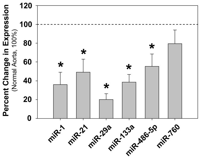
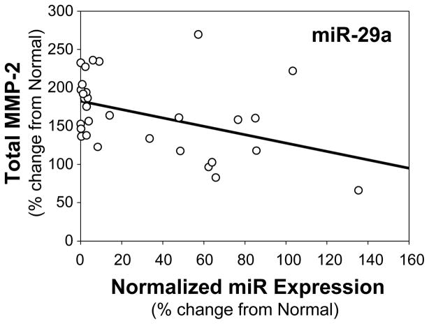
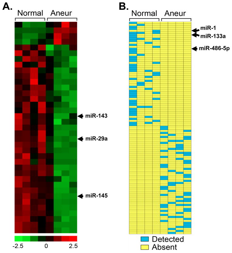
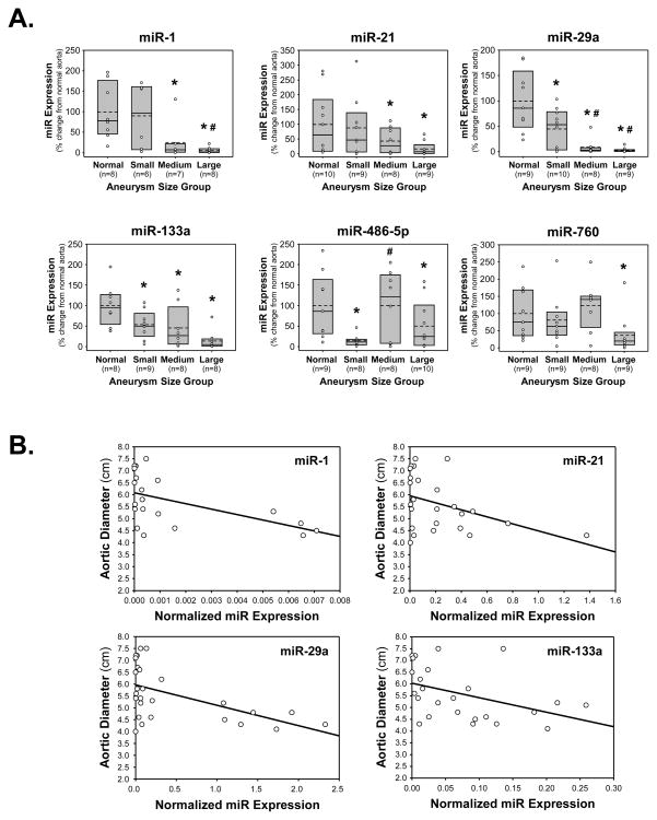
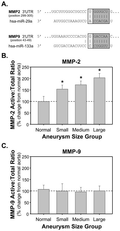
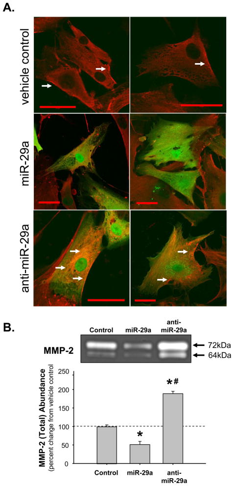
The present study examined miR expression levels in aortic tissue specimens collected from patients with ascending TAAs by quantitative real-time PCR, and observed decreased miR expression (miRs -1, -21, -29a, -133a, and -486) as compared with normal aortic specimens. A significant relationship between miR expression levels (miRs -1, -21, -29a, and -133a) and aortic diameter was identified; as aortic diameter increased, miR expression decreased. Through the use of a bioinformatics approach, members of the matrix metalloproteinase (MMP) family, proteins involved in TAA development, were examined for putative miR binding sites. MMP-2 and MMP-9 were identified as potential targets for miR-29a and miR-133a, respectively, and MMP-2 was subsequently verified as a miR-29a target in vitro. A significant inverse relationship between miR-29a and total MMP-2 was then identified in the clinical TAA specimens.
These findings demonstrate altered miR expression patterns in clinical TAA specimens, suggesting that the loss of specific miR expression may allow for the elaboration of specific MMPs capable of driving aortic remodeling during TAA development. Importantly, these data suggest that these miRs have biological and clinical relevance to the behavior of TAAs and may provide significant targets for therapeutic and diagnostic applications.
越来越多的证据表明,微小RNA(miRNA或miR)表达水平的改变在心血管重塑和疾病进展中起直接作用。尽管miR表达水平的改变已与心脏肥大、纤维化和重塑直接相关,但其在胸主动脉瘤(TAA)发展过程中调节基因表达的作用尚未得到探索。
本研究通过定量实时PCR检测了从升主动脉瘤患者收集的主动脉组织标本中的miR表达水平,发现与正常主动脉标本相比,miR表达水平降低(miR -1、-21、-29a、-133a和-486)。确定了miR表达水平(miR -1、-21、-29a和-133a)与主动脉直径之间存在显著关系;随着主动脉直径增加,miR表达降低。通过生物信息学方法,检查了参与TAA发展的基质金属蛋白酶(MMP)家族成员的假定miR结合位点。MMP-2和MMP-9分别被确定为miR-29a和miR-133a的潜在靶点,随后在体外验证了MMP-2是miR-29a的靶点。然后在临床TAA标本中确定了miR-29a与总MMP-2之间存在显著的负相关关系。
这些发现表明临床TAA标本中miR表达模式发生改变,提示特定miR表达的缺失可能允许在TAA发展过程中产生能够驱动主动脉重塑的特定MMP。重要的是,这些数据表明这些miR与TAA的行为具有生物学和临床相关性,可能为治疗和诊断应用提供重要靶点。