• 文献检索
  • 文档翻译
  • 深度研究
  • 学术资讯
  • Suppr Zotero 插件Zotero 插件
  • 邀请有礼
  • 套餐&价格
  • 历史记录
应用&插件
Suppr Zotero 插件Zotero 插件浏览器插件Mac 客户端Windows 客户端微信小程序
定价
高级版会员购买积分包购买API积分包
服务
文献检索文档翻译深度研究API 文档MCP 服务
关于我们
关于 Suppr公司介绍联系我们用户协议隐私条款
关注我们

Suppr 超能文献

核心技术专利:CN118964589B侵权必究
粤ICP备2023148730 号-1Suppr @ 2026

文献检索

告别复杂PubMed语法,用中文像聊天一样搜索,搜遍4000万医学文献。AI智能推荐,让科研检索更轻松。

立即免费搜索

文件翻译

保留排版,准确专业,支持PDF/Word/PPT等文件格式,支持 12+语言互译。

免费翻译文档

深度研究

AI帮你快速写综述,25分钟生成高质量综述,智能提取关键信息,辅助科研写作。

立即免费体验

基于磁共振成像的膝关节软骨 T2 测量值与膝关节焦点病变和膝关节疼痛的相关性:来自 Osteoarthritis Initiative 的数据。

Association of magnetic resonance imaging-based knee cartilage T2 measurements and focal knee lesions with knee pain: data from the Osteoarthritis Initiative.

机构信息

Department of Radiology and Biomedical Imaging, University of California, San Francisco, CA 94107, USA.

出版信息

Arthritis Care Res (Hoboken). 2012 Feb;64(2):248-55. doi: 10.1002/acr.20672.

DOI:10.1002/acr.20672
PMID:22012846
原文链接:https://pmc.ncbi.nlm.nih.gov/articles/PMC3267009/
Abstract

OBJECTIVE

To evaluate the association of magnetic resonance imaging (MRI)-based knee cartilage T2 measurements and focal knee lesions with knee pain in knees without radiographic osteoarthritis (OA) among subjects with OA risk factors.

METHODS

We studied the right knees of 126 subjects from the Osteoarthritis Initiative database. We randomly selected 42 subjects ages 45-55 years with OA risk factors, right knee pain (Western Ontario and McMaster Universities Osteoarthritis Index [WOMAC] pain score ≥5), no left knee pain (WOMAC pain score 0), and no radiographic OA (Kellgren/Lawrence [K/L] score ≤1) in the right knee. We also selected 2 comparison groups: 42 subjects without knee pain in either knee and 42 with bilateral knee pain. Both groups were frequency matched to subjects with right knee pain only by sex, age, body mass index, and K/L score. All of the subjects underwent 3T MRI of the right knee. Focal knee lesions were assessed and cartilage T2 measurements were performed.

RESULTS

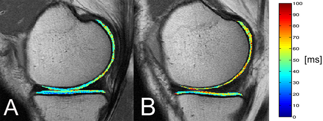
Prevalences of meniscal, bone marrow, and ligamentous lesions and joint effusion were not significantly different between the groups (P > 0.05), while cartilage lesions were more frequent in subjects with right knee pain only compared to subjects without knee pain (P < 0.05). T2 values averaged over all of the compartments were similar in subjects with right knee pain only (mean ± SD 34.4 ± 1.8 msec) and in subjects with bilateral knee pain (mean ± SD 34.7 ± 4.7 msec), but were significantly higher compared to subjects without knee pain (mean ± SD 32.4 ± 1.8 msec; P < 0.05).

CONCLUSION

These results suggest that elevated cartilage T2 values are associated with findings of pain in the early phase of OA, whereas among morphologic knee abnormalities only knee cartilage lesions are significantly associated with knee pain status.

摘要

目的

评估磁共振成像(MRI)膝关节软骨 T2 测量值与膝关节内局灶性病变与存在骨关节炎危险因素的膝关节无放射学骨关节炎(OA)患者膝关节疼痛之间的关系。

方法

我们研究了来自骨关节炎倡议数据库的 126 名受试者的右膝关节。我们随机选择了 42 名年龄在 45-55 岁之间有 OA 危险因素、右膝疼痛(西部安大略省和麦克马斯特大学骨关节炎指数[WOMAC]疼痛评分≥5)、左膝无疼痛(WOMAC 疼痛评分 0)且右膝无放射学 OA(Kellgren/Lawrence [K/L] 评分≤1)的受试者。我们还选择了 2 个对照组:42 名双膝均无疼痛的受试者和 42 名双膝疼痛的受试者。两组均通过性别、年龄、体重指数和 K/L 评分与仅有右膝疼痛的受试者进行频数匹配。所有受试者均接受了右膝关节 3T MRI 检查。评估了局灶性膝关节病变并进行了软骨 T2 测量。

结果

在各组之间,半月板、骨髓、韧带病变和关节积液的患病率无显著差异(P>0.05),而仅有右膝疼痛的受试者的软骨病变较无膝痛的受试者更为常见(P<0.05)。仅有右膝疼痛的受试者(平均±标准差 34.4±1.8msec)和双膝疼痛的受试者(平均±标准差 34.7±4.7msec)的所有节段软骨 T2 值平均值相似,但均显著高于无膝痛的受试者(平均±标准差 32.4±1.8msec;P<0.05)。

结论

这些结果表明,升高的软骨 T2 值与 OA 早期阶段的疼痛表现有关,而在形态学膝关节异常中,仅膝关节软骨病变与膝关节疼痛状态显著相关。

https://cdn.ncbi.nlm.nih.gov/pmc/blobs/66be/3267009/76e684f021cd/nihms332352f1.jpg

相似文献

1
Association of magnetic resonance imaging-based knee cartilage T2 measurements and focal knee lesions with knee pain: data from the Osteoarthritis Initiative.基于磁共振成像的膝关节软骨 T2 测量值与膝关节焦点病变和膝关节疼痛的相关性:来自 Osteoarthritis Initiative 的数据。
Arthritis Care Res (Hoboken). 2012 Feb;64(2):248-55. doi: 10.1002/acr.20672.
2
Correlation of magnetic resonance imaging-based knee cartilage T2 measurements and focal knee lesions with body mass index: thirty-six-month followup data from a longitudinal, observational multicenter study.基于磁共振成像的膝关节软骨 T2 测量值与体质量指数的相关性:一项纵向、观察性多中心研究的 36 个月随访数据。
Arthritis Care Res (Hoboken). 2013 Jan;65(1):23-33. doi: 10.1002/acr.21741.
3
Association of diabetes mellitus and biochemical knee cartilage composition assessed by T relaxation time measurements: Data from the osteoarthritis initiative.糖尿病与 T1 弛豫时间测量评估的膝关节软骨生化成分的相关性:来自骨关节炎倡议的数据。
J Magn Reson Imaging. 2018 Feb;47(2):380-390. doi: 10.1002/jmri.25766. Epub 2017 May 26.
4
Physical activity in relation to knee cartilage T2 progression measured with 3 T MRI over a period of 4 years: data from the Osteoarthritis Initiative.在 4 年的时间里,使用 3T MRI 测量膝关节软骨 T2 进展与身体活动的关系:来自 Osteoarthritis Initiative 的数据。
Osteoarthritis Cartilage. 2013 Oct;21(10):1558-66. doi: 10.1016/j.joca.2013.06.022. Epub 2013 Jul 4.
5
Changes in knee cartilage T2 values over 24 months in subjects with and without risk factors for knee osteoarthritis and their association with focal knee lesions at baseline: data from the osteoarthritis initiative.膝关节软骨 T2 值在 24 个月内的变化,在有和没有膝关节骨关节炎风险因素的受试者中,以及它们与基线时膝关节局灶性病变的关系:来自骨关节炎倡议的数据。
J Magn Reson Imaging. 2012 Feb;35(2):370-8. doi: 10.1002/jmri.22834. Epub 2011 Oct 10.
6
Magnetic resonance imaging lesions are more severe and cartilage T2 relaxation time measurements are higher in isolated lateral compartment radiographic knee osteoarthritis than in isolated medial compartment disease - data from the Osteoarthritis Initiative.来自骨关节炎倡议组织的数据显示,与单纯内侧间室膝关节疾病相比,单纯外侧间室膝关节影像学骨关节炎的磁共振成像病变更严重,软骨T2弛豫时间测量值更高。
Osteoarthritis Cartilage. 2017 Jan;25(1):85-93. doi: 10.1016/j.joca.2016.08.002. Epub 2016 Aug 15.
7
Subjects with higher physical activity levels have more severe focal knee lesions diagnosed with 3T MRI: analysis of a non-symptomatic cohort of the osteoarthritis initiative.体力活动水平较高的受试者在 3T MRI 检查中更易出现严重的膝关节局灶性病变:骨关节炎倡议中无症状队列的分析。
Osteoarthritis Cartilage. 2010 Jun;18(6):776-86. doi: 10.1016/j.joca.2010.02.008. Epub 2010 Feb 14.
8
Association of Superficial Cartilage Transverse Relaxation Time With Osteoarthritis Disease Progression: Data From the Foundation for the National Institutes of Health Biomarker Study of the Osteoarthritis Initiative.超声软骨横向弛豫时间与骨关节炎疾病进展的相关性:来自美国国立卫生研究院生物标志物研究基金会骨关节炎行动计划的数据。
Arthritis Care Res (Hoboken). 2022 Nov;74(11):1888-1893. doi: 10.1002/acr.24627. Epub 2022 Jul 12.
9
Texture analysis of cartilage T2 maps: individuals with risk factors for OA have higher and more heterogeneous knee cartilage MR T2 compared to normal controls--data from the osteoarthritis initiative.软骨 T2 图的纹理分析:与正常对照组相比,有 OA 风险因素的个体的膝关节软骨 MR T2 更高且更不均匀——来自骨关节炎倡议的数据。
Arthritis Res Ther. 2011;13(5):R153. doi: 10.1186/ar3469. Epub 2011 Sep 20.
10
Individual magnetic resonance imaging and radiographic features of knee osteoarthritis in subjects with unilateral knee pain: the health, aging, and body composition study.单侧膝关节疼痛受试者膝骨关节炎的个体磁共振成像和放射学特征:健康、衰老和身体成分研究
Arthritis Rheum. 2012 Oct;64(10):3246-55. doi: 10.1002/art.34594.

引用本文的文献

1
Pharmacist-Driven Chondroprotection in Osteoarthritis: A Multifaceted Approach Using Patient Education, Information Visualization, and Lifestyle Integration.药剂师主导的骨关节炎软骨保护:一种采用患者教育、信息可视化和生活方式整合的多方面方法。
Pharmacy (Basel). 2025 Aug 1;13(4):106. doi: 10.3390/pharmacy13040106.
2
Assessment of articular cartilage degradation in response to an impact injury using µMRI.使用 µMRI 评估关节软骨对撞击损伤的降解。
Connect Tissue Res. 2024 Mar;65(2):146-160. doi: 10.1080/03008207.2024.2319050. Epub 2024 Feb 28.
3
Quantitative Evaluation of Knee Cartilage in Professional Martial Arts Athletes Using T2 Mapping: A Comparative Study.

本文引用的文献

1
Physical activity is associated with magnetic resonance imaging-based knee cartilage T2 measurements in asymptomatic subjects with and those without osteoarthritis risk factors.在有和没有骨关节炎风险因素的无症状受试者中,身体活动与基于磁共振成像的膝关节软骨T2测量值相关。
Arthritis Rheum. 2011 Aug;63(8):2248-56. doi: 10.1002/art.30419.
2
A novel fast knee cartilage segmentation technique for T2 measurements at MR imaging--data from the Osteoarthritis Initiative.一种用于磁共振成像 T2 测量的新型快速膝关节软骨分割技术——来自骨关节炎倡议的数据。
Osteoarthritis Cartilage. 2011 Aug;19(8):984-9. doi: 10.1016/j.joca.2011.04.002. Epub 2011 Apr 12.
3
基于 T2 图谱的专业武术运动员膝关节软骨定量评估:一项对比研究。
J Athl Train. 2024 Oct 1;59(10):1012-1018. doi: 10.4085/1062-6050-0127.23.
4
MRI-based T and T cartilage compositional imaging in osteoarthritis: what have we learned and what is needed to apply it clinically and in a trial setting?基于 MRI 的 T1 和 T2 软骨成分成像在骨关节炎中的应用:我们已经学到了什么,以及在临床和试验环境中应用它需要什么?
Skeletal Radiol. 2023 Nov;52(11):2137-2147. doi: 10.1007/s00256-023-04310-x. Epub 2023 Mar 31.
5
Osteophytes mediate the associations between cartilage morphology and changes in knee symptoms in patients with knee osteoarthritis.骨赘介导了膝关节骨关节炎患者软骨形态与膝关节症状变化之间的关联。
Arthritis Res Ther. 2022 Sep 8;24(1):217. doi: 10.1186/s13075-022-02905-8.
6
Sandwich Technique for Large Osteochondral Lesions of the Knee.膝关节大块骨软骨病变的三明治技术。
Cartilage. 2022 Jul-Sep;13(3):19476035221102571. doi: 10.1177/19476035221102571.
7
The Effect of Lesion Size on Pain and Function in Patients Scheduled for Cartilage Surgery of the Knee.膝关节软骨手术患者的病变大小对疼痛和功能的影响。
Cartilage. 2022 Apr-Jun;13(2):19476035221109242. doi: 10.1177/19476035221109242.
8
Meniscal anterior and posterior horn heights are associated with MRI-defined knee structural abnormalities in middle-aged and elderly patients with symptomatic knee osteoarthritis.前、后半月板角高度与中老年膝关节骨关节炎患者膝关节结构异常的 MRI 表现有关。
BMC Musculoskelet Disord. 2022 Mar 8;23(1):218. doi: 10.1186/s12891-022-05143-w.
9
Machine learning to predict incident radiographic knee osteoarthritis over 8 Years using combined MR imaging features, demographics, and clinical factors: data from the Osteoarthritis Initiative.机器学习预测 8 年内膝关节骨关节炎的影像学表现:来自骨关节炎倡议的联合磁共振成像特征、人口统计学和临床因素的数据。
Osteoarthritis Cartilage. 2022 Feb;30(2):270-279. doi: 10.1016/j.joca.2021.11.007. Epub 2021 Nov 18.
10
Uncovering associations between data-driven learned qMRI biomarkers and chronic pain.揭示数据驱动的学习 qMRI 生物标志物与慢性疼痛之间的关联。
Sci Rep. 2021 Nov 9;11(1):21989. doi: 10.1038/s41598-021-01111-x.
Vastus lateralis/vastus medialis cross-sectional area ratio impacts presence and degree of knee joint abnormalities and cartilage T2 determined with 3T MRI - an analysis from the incidence cohort of the Osteoarthritis Initiative.
股外侧肌/股内侧肌横截面积比影响膝关节异常和软骨 T2 的存在和程度,这是使用 3T MRI 从 Osteoarthritis Initiative 发病队列分析得出的结果。
Osteoarthritis Cartilage. 2011 Jan;19(1):65-73. doi: 10.1016/j.joca.2010.10.023. Epub 2010 Oct 31.
4
Cartilage and meniscus assessment using T1rho and T2 measurements in healthy subjects and patients with osteoarthritis.在健康受试者和骨关节炎患者中使用 T1rho 和 T2 测量评估软骨和半月板。
Osteoarthritis Cartilage. 2010 Nov;18(11):1408-16. doi: 10.1016/j.joca.2010.07.012. Epub 2010 Aug 7.
5
Relation of synovitis to knee pain using contrast-enhanced MRIs.使用对比增强 MRI 评估滑膜炎与膝关节疼痛的关系。
Ann Rheum Dis. 2010 Oct;69(10):1779-83. doi: 10.1136/ard.2009.121426. Epub 2010 May 14.
6
Subjects with higher physical activity levels have more severe focal knee lesions diagnosed with 3T MRI: analysis of a non-symptomatic cohort of the osteoarthritis initiative.体力活动水平较高的受试者在 3T MRI 检查中更易出现严重的膝关节局灶性病变:骨关节炎倡议中无症状队列的分析。
Osteoarthritis Cartilage. 2010 Jun;18(6):776-86. doi: 10.1016/j.joca.2010.02.008. Epub 2010 Feb 14.
7
Patellar cartilage: T2 values and morphologic abnormalities at 3.0-T MR imaging in relation to physical activity in asymptomatic subjects from the osteoarthritis initiative.髌软骨:3.0T MRI 成像中与无症状受试者体力活动相关的 T2 值和形态异常——来自骨关节炎倡议研究。
Radiology. 2010 Feb;254(2):509-20. doi: 10.1148/radiol.09090596. Epub 2009 Dec 17.
8
Does joint alignment affect the T2 values of cartilage in patients with knee osteoarthritis?关节对线是否会影响膝骨关节炎患者软骨的 T2 值?
Eur Radiol. 2010 Jun;20(6):1532-8. doi: 10.1007/s00330-009-1689-7. Epub 2009 Dec 16.
9
Denuded subchondral bone and knee pain in persons with knee osteoarthritis.膝关节骨关节炎患者的软骨下骨裸露与膝关节疼痛。
Arthritis Rheum. 2009 Dec;60(12):3703-10. doi: 10.1002/art.25014.
10
Magnetic resonance imaging-based semiquantitative and quantitative assessment in osteoarthritis.基于磁共振成像的骨关节炎半定量和定量评估。
Rheum Dis Clin North Am. 2009 Aug;35(3):521-55. doi: 10.1016/j.rdc.2009.08.006.