Transcranial Magnetic Stimulation Laboratory, Center for Mind/Brain Sciences, University of Trento Trento, Italy.
Front Neural Circuits. 2011 Oct 18;5:14. doi: 10.3389/fncir.2011.00014. eCollection 2011.
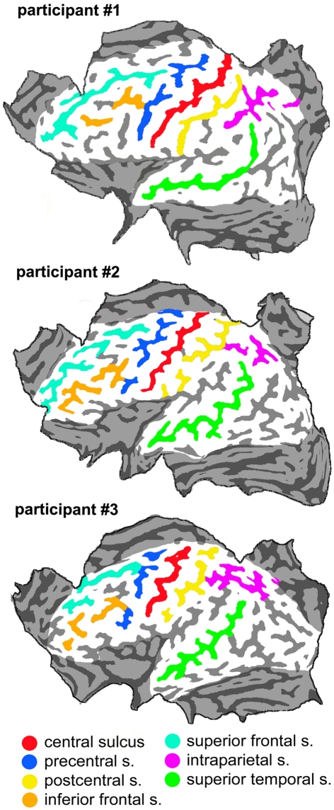
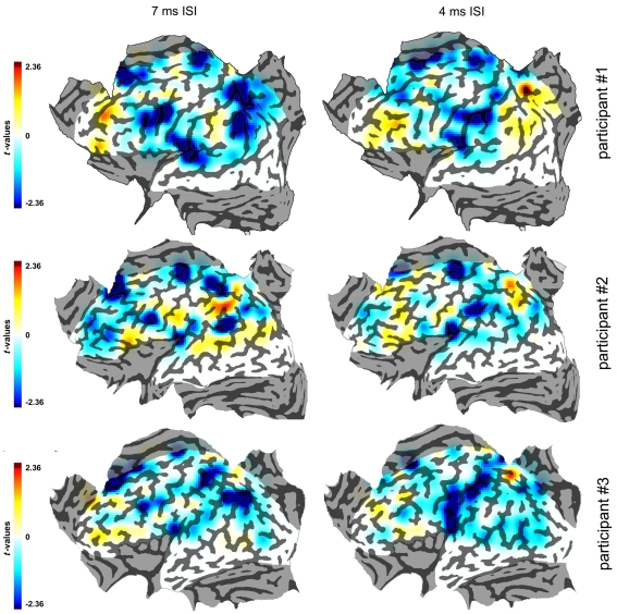
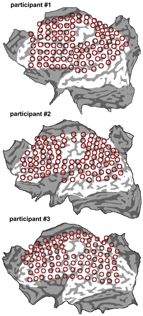
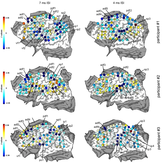
Skilled hand function relies heavily on the integrity of the primary motor cortex (M1) and on a web of cortico-cortical connections projecting onto it. We used a novel explorative paradigm to map the origin of cortico-M1 pathways assessed by dual transcranial magnetic stimulation (TMS) in three healthy participants. Subthreshold conditioning TMS (cTMS) was delivered over a grid of ≈100 spots. Covering the left hemisphere, and was followed by suprathreshold test (tTMS) delivered over the ipsilateral M1. Grid points were tested eight times, with inter-stimulus intervals between cTMS and tTMS of 4 and 7 ms. Participants were asked to stay relaxed with no particular task. Motor evoked potentials (MEPs) from cTMS + tTMS were normalized to MEPs from tTMS alone and were compared to the value expected from tTMS alone using t-statistics. The t-values from each grid point were then used to plot statistical maps. Several foci of significant cortico-M1 interactions were found in the dorsal-medial frontal cortex, in the ventral frontal cortex, in the superior and inferior parietal lobules and in the parietal operculum. The majority of active foci had inhibitory effects on corticospinal excitability. The spatial location of the network of different subjects overlapped but with some anatomical variation of single foci. TMS statistical mapping during the resting state revealed a complex inhibitory cortical network. The explorative approach to TMS as a brain mapping tool produced results that are self-standing in single subjects overcoming inter-individual variability of cortical active sites.
熟练的手部功能依赖于初级运动皮层(M1)的完整性,以及投射到其上的皮质-皮质连接网络。我们使用一种新的探索性范式,在三名健康参与者中通过双经颅磁刺激(TMS)来绘制皮质-M1 通路的起源图。阈下条件 TMS(cTMS)在大约 100 个点的网格上进行。覆盖左半球,并随后在同侧 M1 上进行超阈值测试(tTMS)。网格点测试了八次,cTMS 和 tTMS 之间的刺激间隔为 4 和 7ms。参与者被要求保持放松,没有特定的任务。cTMS+tTMS 的运动诱发电位(MEP)被归一化为单独的 tTMS 的 MEP,并使用 t 统计量与单独的 tTMS 的预期值进行比较。然后,使用每个网格点的 t 值绘制统计图。在背内侧额皮质、腹侧额皮质、顶叶上、下小叶和顶叶外侧区发现了几个有意义的皮质-M1 相互作用的焦点。大多数活跃的焦点对皮质脊髓兴奋性有抑制作用。不同受试者的网络的空间位置重叠,但单个焦点的解剖变异。静息状态下 TMS 的统计映射揭示了一个复杂的抑制性皮质网络。TMS 作为大脑映射工具的探索性方法产生了独立于单个受试者的结果,克服了皮质活性部位的个体间变异性。