• 文献检索
  • 文档翻译
  • 深度研究
  • 学术资讯
  • Suppr Zotero 插件Zotero 插件
  • 邀请有礼
  • 套餐&价格
  • 历史记录
应用&插件
Suppr Zotero 插件Zotero 插件浏览器插件Mac 客户端Windows 客户端微信小程序
定价
高级版会员购买积分包购买API积分包
服务
文献检索文档翻译深度研究API 文档MCP 服务
关于我们
关于 Suppr公司介绍联系我们用户协议隐私条款
关注我们

Suppr 超能文献

核心技术专利:CN118964589B侵权必究
粤ICP备2023148730 号-1Suppr @ 2026

文献检索

告别复杂PubMed语法,用中文像聊天一样搜索,搜遍4000万医学文献。AI智能推荐,让科研检索更轻松。

立即免费搜索

文件翻译

保留排版,准确专业,支持PDF/Word/PPT等文件格式,支持 12+语言互译。

免费翻译文档

深度研究

AI帮你快速写综述,25分钟生成高质量综述,智能提取关键信息,辅助科研写作。

立即免费体验

人类下颌骨生长的三维容积评估

3-d volumetric evaluation of human mandibular growth.

作者信息

Reynolds Mathew, Reynolds Michael, Adeeb Samer, El-Bialy Tarek

机构信息

Department of Civil and Environmental Engineering, University of Alberta, Edmonton, AB, Canada.

出版信息

Open Biomed Eng J. 2011;5:83-9. doi: 10.2174/1874120701105010083. Epub 2011 Oct 14.

DOI:10.2174/1874120701105010083
PMID:22046201
原文链接:https://pmc.ncbi.nlm.nih.gov/articles/PMC3204416/
Abstract

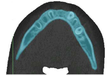
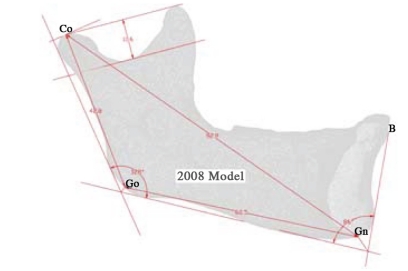
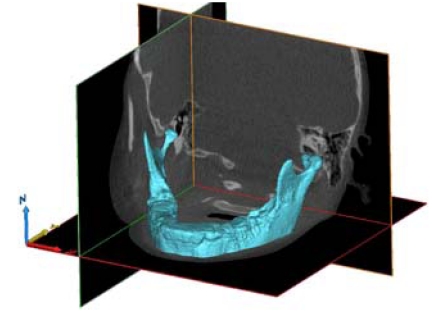
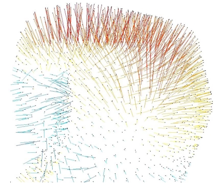
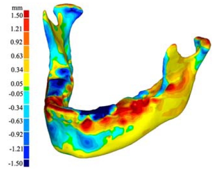
Bone growth is a complex process that is controlled by a multitude of mechanisms that are not fully understood.Most of the current methods employed to measure the growth of bones focus on either studying cadaveric bones from different individuals of different ages, or successive two-dimensional (2D) radiographs. Both techniques have their known limitations. The purpose of this study was to explore a technique for quantifying the three dimensional (3D) growth of an adolescent human mandible over the period of one year utilizing cone beam computed tomography (CBCT) scans taken for regular orthodontic records. Three -dimensional virtual models were created from the CBCT data using mainstream medical imaging software. A comparison between computer-generated surface meshes of successive 3-D virtual models illustrates the magnitude of relative mandible growth. The results of this work are in agreement with previously reported data from human cadaveric studies and implantable marker studies. The presented method provides a new relatively simple basis (utilizing commercially available software) to visualize and evaluate individualized 3D (mandibular) growth in vivo.

摘要

骨骼生长是一个复杂的过程,受多种尚未完全明确的机制控制。目前用于测量骨骼生长的大多数方法要么侧重于研究不同年龄个体的尸体骨骼,要么侧重于连续的二维(2D)X光片。这两种技术都有其已知的局限性。本研究的目的是探索一种技术,利用为常规正畸记录拍摄的锥形束计算机断层扫描(CBCT),对青少年人类下颌骨在一年时间内的三维(3D)生长进行量化。使用主流医学成像软件从CBCT数据创建三维虚拟模型。连续三维虚拟模型的计算机生成表面网格之间的比较说明了下颌骨相对生长的幅度。这项工作的结果与先前来自人体尸体研究和可植入标记物研究报告的数据一致。所提出的方法提供了一个新的相对简单的基础(利用商业软件),以在体内可视化和评估个性化的三维(下颌骨)生长。

https://cdn.ncbi.nlm.nih.gov/pmc/blobs/8aac/3204416/3dafd0de5a5c/TOBEJ-5-83_F8.jpg
https://cdn.ncbi.nlm.nih.gov/pmc/blobs/8aac/3204416/b0e9b831a4d8/TOBEJ-5-83_F1.jpg
https://cdn.ncbi.nlm.nih.gov/pmc/blobs/8aac/3204416/6c9db94faa12/TOBEJ-5-83_F2.jpg
https://cdn.ncbi.nlm.nih.gov/pmc/blobs/8aac/3204416/44a69f93f7cd/TOBEJ-5-83_F3.jpg
https://cdn.ncbi.nlm.nih.gov/pmc/blobs/8aac/3204416/9958c244ca44/TOBEJ-5-83_F4.jpg
https://cdn.ncbi.nlm.nih.gov/pmc/blobs/8aac/3204416/15892c0a1cdc/TOBEJ-5-83_F5.jpg
https://cdn.ncbi.nlm.nih.gov/pmc/blobs/8aac/3204416/7e68a793178b/TOBEJ-5-83_F6.jpg
https://cdn.ncbi.nlm.nih.gov/pmc/blobs/8aac/3204416/da9a1c1f8e02/TOBEJ-5-83_F7.jpg
https://cdn.ncbi.nlm.nih.gov/pmc/blobs/8aac/3204416/3dafd0de5a5c/TOBEJ-5-83_F8.jpg

相似文献

1
3-d volumetric evaluation of human mandibular growth.人类下颌骨生长的三维容积评估
Open Biomed Eng J. 2011;5:83-9. doi: 10.2174/1874120701105010083. Epub 2011 Oct 14.
2
The impact of different cone beam computed tomography and multi-slice computed tomography scan parameters on virtual three-dimensional model accuracy using a highly precise ex vivo evaluation method.使用高精度体外评估方法,不同锥束计算机断层扫描和多层螺旋计算机断层扫描参数对虚拟三维模型准确性的影响。
J Craniomaxillofac Surg. 2016 May;44(5):632-6. doi: 10.1016/j.jcms.2016.02.005. Epub 2016 Feb 13.
3
A comparison between two-dimensional and three-dimensional cephalometry on frontal radiographs and on cone beam computed tomography scans of human skulls.人类颅骨正位X线片和锥形束计算机断层扫描上二维与三维头影测量的比较。
Eur J Oral Sci. 2009 Jun;117(3):300-5. doi: 10.1111/j.1600-0722.2009.00633.x.
4
Monitoring of typodont root movement via crown superimposition of single cone-beam computed tomography and consecutive intraoral scans.通过单锥束 CT 牙冠重叠和连续口内扫描监测牙模根移动。
Am J Orthod Dentofacial Orthop. 2014 Mar;145(3):399-409. doi: 10.1016/j.ajodo.2013.12.011.
5
[Exploring a new method for superimposition of pre-treatment and post-treatment mandibular digital dental casts in adults].[探索成人治疗前与治疗后下颌数字化牙模叠加的新方法]
Beijing Da Xue Xue Bao Yi Xue Ban. 2018 Apr 18;50(2):271-278.
6
One Step before 3D Printing-Evaluation of Imaging Software Accuracy for 3-Dimensional Analysis of the Mandible: A Comparative Study Using a Surface-to-Surface Matching Technique.3D打印前的一步——下颌骨三维分析成像软件准确性评估:一项使用表面到表面匹配技术的比较研究
Materials (Basel). 2020 Jun 21;13(12):2798. doi: 10.3390/ma13122798.
7
Evaluation of mandibular growth using cone-beam computed tomography in a rabbit model: a pilot study.使用锥形束计算机断层扫描评估兔模型中的下颌骨生长:一项初步研究。
N Z Dent J. 2012 Mar;108(1):9-12.
8
Volumetric Evaluation of Safe Zone for Bone Harvesting From Symphysis Region by Using Cone Beam Computed Tomography.使用锥形束计算机断层扫描对耻骨联合区域取骨安全区进行容积评估
Implant Dent. 2016 Dec;25(6):758-761. doi: 10.1097/ID.0000000000000450.
9
Superimposition of 3-dimensional cone-beam computed tomography models of growing patients.生长发育期患者的三维锥形束计算机断层扫描模型的叠加
Am J Orthod Dentofacial Orthop. 2009 Jul;136(1):94-9. doi: 10.1016/j.ajodo.2009.01.018.
10
Determining the Accuracy of the Mandibular Canal Region in 3D Biomodels Fabricated from CBCT Scanned Data: A Cadaveric Study.确定由CBCT扫描数据制作的三维生物模型中下颌管区域的准确性:一项尸体研究。
Curr Med Imaging Rev. 2019;15(7):645-653. doi: 10.2174/1573405614666181012144745.

引用本文的文献

1
Comparison of Nasal Analysis by Photographs (2D) against Low-cost Surface Laser Imaging (3D) and against Computed Axial Tomography Imaging.通过照片(二维)进行的鼻腔分析与低成本表面激光成像(三维)及计算机轴向断层扫描成像的比较。
Indian J Plast Surg. 2022 Dec 16;56(2):147-152. doi: 10.1055/s-0042-1759724. eCollection 2023 Apr.
2
Datasets describing the morphometric evaluation of the adult rat mandible following growth stimulation of the condyle using ultrasound.描述使用超声刺激髁突后成年大鼠下颌骨形态计量学评估的数据集。
Data Brief. 2022 Oct 8;45:108664. doi: 10.1016/j.dib.2022.108664. eCollection 2022 Dec.
3
A three-dimensional statistical shape model of the growing mandible.

本文引用的文献

1
3D printing based on imaging data: review of medical applications.基于成像数据的 3D 打印:医学应用综述。
Int J Comput Assist Radiol Surg. 2010 Jul;5(4):335-41. doi: 10.1007/s11548-010-0476-x. Epub 2010 May 15.
2
Evaluating the mandible with cone-beam computed tomography.应用锥形束 CT 评估下颌骨
Am J Orthod Dentofacial Orthop. 2010 Apr;137(4 Suppl):S58-70. doi: 10.1016/j.ajodo.2009.01.025.
3
Mandibular growth, remodeling, and maturation during infancy and early childhood.下颌骨在婴儿期和幼儿期的生长、重塑和成熟。
生长下颌骨的三维统计形状模型。
Sci Rep. 2021 Sep 22;11(1):18843. doi: 10.1038/s41598-021-98421-x.
4
The effect of low intensity pulsed ultrasound on mandibular condylar growth in young adult rats.低强度脉冲超声对年轻成年大鼠下颌髁突生长的影响。
Bone Rep. 2021 Sep 3;15:101122. doi: 10.1016/j.bonr.2021.101122. eCollection 2021 Dec.
5
3D Printing-Encompassing the Facets of Dentistry.3D打印——涵盖牙科的各个方面。
Front Bioeng Biotechnol. 2018 Nov 22;6:172. doi: 10.3389/fbioe.2018.00172. eCollection 2018.
6
Dimensional Error in Rapid Prototyping with Open Source Software and Low-cost 3D-printer.使用开源软件和低成本3D打印机进行快速成型时的尺寸误差
Plast Reconstr Surg Glob Open. 2018 Jan 25;6(1):e1646. doi: 10.1097/GOX.0000000000001646. eCollection 2018 Jan.
7
A Novel Registration-Based Semiautomatic Mandible Segmentation Pipeline Using Computed Tomography Images to Study Mandibular Development.一种基于配准的新型半自动下颌骨分割流程,使用计算机断层扫描图像研究下颌骨发育。
J Comput Assist Tomogr. 2018 Mar/Apr;42(2):306-316. doi: 10.1097/RCT.0000000000000669.
8
Comparison of measurements of mandible growth using cone beam computed tomography and its synthesized cephalograms.使用锥形束计算机断层扫描及其合成头影测量片对下颌骨生长测量的比较。
Biomed Eng Online. 2014 Sep 10;13:133. doi: 10.1186/1475-925X-13-133.
Angle Orthod. 2010 Jan;80(1):97-105. doi: 10.2319/020309-67.1.
4
An experimental method for stereolithic mandible fabrication and image preparation.一种用于立体光刻下颌骨制造和图像制备的实验方法。
Open Biomed Eng J. 2007 Jul 17;1:4-10. doi: 10.2174/1874120700701010004.
5
Effects of mastication on mandibular growth evaluated by microcomputed tomography.通过微计算机断层扫描评估咀嚼对下颌生长的影响。
Eur J Orthod. 2010 Feb;32(1):66-70. doi: 10.1093/ejo/cjp060. Epub 2009 Jul 31.
6
Simulation of biological growth.生物生长模拟
Comput Methods Biomech Biomed Engin. 2009 Dec;12(6):617-26. doi: 10.1080/10255840902802909.
7
Comparison of relative mandibular growth vectors with high-resolution 3-dimensional imaging.利用高分辨率三维成像技术比较下颌骨相对生长向量
Am J Orthod Dentofacial Orthop. 2005 Jul;128(1):27-34. doi: 10.1016/j.ajodo.2004.03.033.
8
Assessment of mandibular growth and response to orthopedic treatment with 3-dimensional magnetic resonance images.使用三维磁共振成像评估下颌骨生长及对正畸治疗的反应。
Am J Orthod Dentofacial Orthop. 2005 Jul;128(1):16-26. doi: 10.1016/j.ajodo.2004.03.032.
9
Three-dimensional simulation and prediction of craniofacial surgery.颅面外科手术的三维模拟与预测
Orthod Craniofac Res. 2003;6 Suppl 1:102-7. doi: 10.1034/j.1600-0544.2003.242.x.
10
Growth modeling of human mandibles using non-Euclidean metrics.使用非欧几里得度量对人类下颌骨进行生长建模。
Med Image Anal. 2003 Dec;7(4):425-33. doi: 10.1016/s1361-8415(03)00034-3.