Department of Surgery, Hanyang University College of Medicine, Seoul, Korea.
J Gastric Cancer. 2011 Mar;11(1):38-45. doi: 10.5230/jgc.2011.11.1.38. Epub 2011 Mar 31.
Bone metastasis from stomach cancer occurs only rarely and it is known to have a very poor prognosis. This study examined the clinical characteristics and prognosis of patients who were diagnosed with stomach cancer and bone metastasis.
The subjects were 19 patients who were diagnosed with stomach cancer at Hanyang University Medical Center from June 1992 to August 2010 and they also had bone metastasis. The survival rate according to many clinicopathologic factors was retrospectively analyzed.
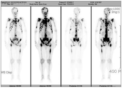
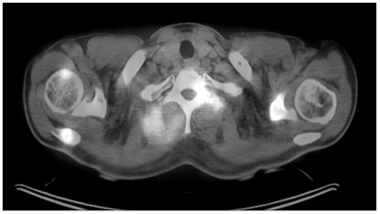
11 patients out of 18 patients (61%) who received an operation were in stage IV and the most common bone metastasis location was the spine. Bone scintigraphy was mostly used for diagnosing bone metastasis and PET-CT and magnetic resonance imaging were used singly or together. The serum alkaline phosphatase at the time of diagnosis had increased in 12 cases and there were clinical symptoms (bone pain) in 16 cases. Treatment was given to 14 cases and it was mostly radiotherapy. There were 2 cases of discovering bone metastasis at the time of diagnosing stomach cancer. The interval after operation to the time of diagnosing bone metastasis for the 18 cases that received a stomach cancer operation was on average 14.9±17.3 months and the period until death after the diagnosis of bone metastasis was on average 3.8±2.6 months. As a result of univariate survival rate analysis, the group that was treated for bone metastasis had a significantly better survival period when the bone metastasis was singular rather than multiple, as compared to the non-treatment group, yet both factors were not independent prognosis factors on multivariate survival analysis.
An examination to confirm the status of bone metastasis when conducting a radio-tracer test after the initial diagnosis and also after an operation is needed for stomach cancer patients, and bone scintigraphy is the most helpfully modality. Making the diagnosis at the early stage and suitable treatments are expected to enhance the survival rate and improve the quality of life even for the patients with bone metastasis.
胃癌骨转移极为罕见,且预后极差。本研究旨在探讨诊断为胃癌伴骨转移患者的临床特征及预后。
回顾性分析 1992 年 6 月至 2010 年 8 月在汉阳大学医疗中心诊断为胃癌且发生骨转移的 19 例患者的临床病理资料,分析患者的生存率与多种临床病理因素的关系。
18 例行手术治疗的患者中 11 例(61%)为Ⅳ期,最常见的骨转移部位为脊柱。骨闪烁扫描是诊断骨转移的常用方法,而单独或联合应用 PET-CT 和磁共振成像检查的病例也较多。12 例患者在诊断时碱性磷酸酶升高,16 例患者出现骨痛等临床症状。14 例患者接受了治疗,其中以放疗为主。2 例患者在诊断胃癌时即发现骨转移。18 例行胃癌手术的患者中,术后至诊断骨转移的平均时间为 14.9±17.3 个月,诊断骨转移后至死亡的平均时间为 3.8±2.6 个月。单因素生存分析显示,与未治疗组相比,单发骨转移患者接受骨转移治疗的生存时间明显更长,但这两个因素在多因素生存分析中均不是独立的预后因素。
胃癌患者在初始诊断及术后进行放射性核素检查时,应进行骨转移状态检查,骨闪烁扫描是最有帮助的检查方法。早期诊断和适当的治疗有望提高生存率,改善生活质量,即使是发生骨转移的患者也不例外。