Academic Oncology, University of Nottingham, School of Molecular Medical Sciences, Nottingham University Hospitals NHS Trust, City Hospital Campus, Nottingham, UK.
Mod Pathol. 2012 Apr;25(4):493-504. doi: 10.1038/modpathol.2011.182. Epub 2011 Nov 11.
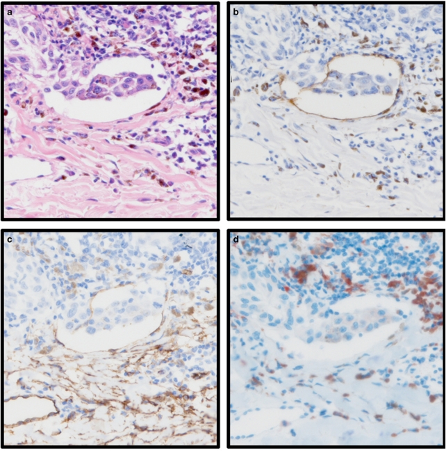
The aims of this study were to investigate the role of vascular invasion (blood and lymphatic), vessel density and the presence of tumour-associated macrophages as prognostic markers in 202 cutaneous melanoma patients. Sections of primary melanoma were stained with lymphatic-specific antibody D2-40 to assess lymphatic vessel invasion and density in intratumoural and peritumoural areas; an antibody against endothelial marker CD34 was used to determine blood vessel invasion and density, and an antibody against CD68 was used to determine macrophage counts. Immunohistochemically determined vascular invasion (combined blood and lymphatic) was compared with that determined using haematoxylin and eosin (H&E) staining. The use of immunohistochemistry increased detection of vascular invasion from 8-30% of patients, and histological exam of H&E-stained tissue was associated with a false positive rate of 64%. Lymphatic vessel invasion occurred at a much higher frequency than blood vessel invasion (27 and 4% of patients, respectively). Although immunohistochemically detected vessel invasion was significantly associated with histological markers of adverse prognosis, such as increased Breslow thickness, ulceration and mitotic rate (all P<0.001), no associations with relapse-free or overall survival were observed. High macrophage counts were significantly associated with markers of aggressive disease, such as Breslow thickness, ulceration and mitotic rate (P<0.001, P<0.001, P=0.005, respectively), and lymphatic vessel invasion and high microvessel density (P=0.002 and P=0.003, respectively). These results suggest that vascular invasion is more accurately detected using immunohistochemistry and occurs predominantly via lymphatic vessels. The association of vessel characteristics with histological characteristics of the primary melanoma provides evidence for their biological importance in melanoma, but that they were not associated with clinical outcome attests to the value of existing histological prognostic biomarkers. We note that a high macrophage count may be associated with neovascularisation and primary tumour growth, and may also promote invasion through lymphatic vessels.
本研究的目的是探究血管侵犯(血道和淋巴道)、血管密度以及肿瘤相关巨噬细胞作为 202 例皮肤黑色素瘤患者预后标志物的作用。对原发黑色素瘤切片进行淋巴管特异性抗体 D2-40 染色,以评估肿瘤内和肿瘤周围区域的淋巴道侵犯和密度;使用内皮标志物 CD34 抗体来确定血管侵犯和密度,并用 CD68 抗体来确定巨噬细胞计数。通过免疫组织化学确定的血管侵犯(血道和淋巴道联合)与苏木精和伊红(H&E)染色确定的血管侵犯进行了比较。免疫组织化学检测方法增加了血管侵犯的检出率,从 8%至 30%的患者中检出,而 H&E 染色组织的组织学检查与 64%的假阳性率相关。淋巴道侵犯的发生率远高于血管侵犯(分别为 27%和 4%的患者)。尽管免疫组织化学检测到的血管侵犯与不良预后的组织学标志物显著相关,如增加的 Breslow 厚度、溃疡和有丝分裂率(均 P<0.001),但与无复发生存或总生存无相关性。高巨噬细胞计数与侵袭性疾病标志物显著相关,如 Breslow 厚度、溃疡和有丝分裂率(P<0.001,P<0.001,P=0.005),以及淋巴道侵犯和高微血管密度(P=0.002 和 P=0.003)。这些结果表明,免疫组织化学检测血管侵犯更为准确,且主要通过淋巴道发生。血管特征与原发黑色素瘤的组织学特征相关,这为它们在黑色素瘤中的生物学重要性提供了证据,但它们与临床结局无关,这证明了现有组织学预后生物标志物的价值。我们注意到,高巨噬细胞计数可能与新生血管形成和原发肿瘤生长有关,也可能通过淋巴道促进侵犯。