• 文献检索
  • 文档翻译
  • 深度研究
  • 学术资讯
  • Suppr Zotero 插件Zotero 插件
  • 邀请有礼
  • 套餐&价格
  • 历史记录
应用&插件
Suppr Zotero 插件Zotero 插件浏览器插件Mac 客户端Windows 客户端微信小程序
定价
高级版会员购买积分包购买API积分包
服务
文献检索文档翻译深度研究API 文档MCP 服务
关于我们
关于 Suppr公司介绍联系我们用户协议隐私条款
关注我们

Suppr 超能文献

核心技术专利:CN118964589B侵权必究
粤ICP备2023148730 号-1Suppr @ 2026

文献检索

告别复杂PubMed语法,用中文像聊天一样搜索,搜遍4000万医学文献。AI智能推荐,让科研检索更轻松。

立即免费搜索

文件翻译

保留排版,准确专业,支持PDF/Word/PPT等文件格式,支持 12+语言互译。

免费翻译文档

深度研究

AI帮你快速写综述,25分钟生成高质量综述,智能提取关键信息,辅助科研写作。

立即免费体验

与圆顶状黄斑和倾斜视盘相关的浆液性黄斑脱离。

Serous macular detachment associated with dome-shaped macula and tilted disc.

作者信息

Pardo-López Diamar, Gallego-Pinazo Roberto, Mateo Carlos, Rohrweck Stephanie, Suelves Ana M, Dolz-Marco Rosa, Mullor José Luis, Díaz-Llopis Manuel

机构信息

Department of Ophthalmology, University and Polytechnic Hospital La Fe, Barcelona, Spain.

出版信息

Case Rep Ophthalmol. 2011 Jan;2(1):111-5. doi: 10.1159/000326918. Epub 2011 Mar 16.

DOI:10.1159/000326918
PMID:22087101
原文链接:https://pmc.ncbi.nlm.nih.gov/articles/PMC3214892/
Abstract

INTRODUCTION

An entirely new type of staphyloma has been recently described as dome-shaped macula (DSM). It is characterized by an abnormal convex macular contour within the concavity of a posterior staphyloma. We found DSM associated with serous macular detachment (SMD) and tilted disc in two consecutive cases.

CASE REPORTS

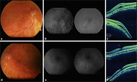
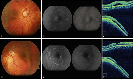
Case 1: A 37-year-old female presented to our department because of sudden onset blurred vision in her right eye (OD). The best-corrected visual acuity (BCVA) was 0.5 in both eyes. Funduscopy evidenced bilateral tilted disc associated with posterior staphyloma. Optical coherence tomography (OCT) demonstrated a DSM with SMD in her OD. After 15 months of follow-up, BCVA of her OD remained stable with chronic SMD. Case 2: A 32-year-old female presented to our department because of blurred vision in her OD. The BCVA was 0.4 in the OD and 1.0 in the left eye (OS). Bilateral tilted disc and posterior staphyloma were evidenced in the funduscopy. OCT demonstrated a bilateral DSM with SMD in her OD. After 45 months of follow-up, two further episodes of transient SMD were observed in her OD and seven in her OS. The final BCVA was 0.63 in the OD and 0.8 in the OS.

DISCUSSION

SMD associated with tilted disc constitutes a potential cause of subretinal fluid accumulation in myopic patients. OCT is essential for the detection of both SMD and DSM.

摘要

引言

最近一种全新类型的葡萄肿被描述为穹窿状黄斑(DSM)。其特征是在后葡萄肿凹陷内黄斑轮廓异常凸起。我们在连续两例病例中发现DSM与浆液性黄斑脱离(SMD)和倾斜视盘相关。

病例报告

病例1:一名37岁女性因右眼突然视力模糊就诊于我科。双眼最佳矫正视力(BCVA)均为0.5。眼底检查发现双侧倾斜视盘伴后葡萄肿。光学相干断层扫描(OCT)显示其右眼存在伴有SMD的DSM。随访15个月后,其右眼BCVA随慢性SMD保持稳定。病例2:一名32岁女性因右眼视力模糊就诊于我科。右眼BCVA为0.4,左眼(OS)为1.0。眼底检查发现双侧倾斜视盘和后葡萄肿。OCT显示其右眼存在双侧伴有SMD的DSM。随访45个月后,观察到其右眼又出现了两次短暂性SMD,左眼出现了七次。最终右眼BCVA为0.63,左眼为0.8。

讨论

与倾斜视盘相关的SMD是近视患者视网膜下液体积聚的一个潜在原因。OCT对于检测SMD和DSM至关重要。

https://cdn.ncbi.nlm.nih.gov/pmc/blobs/a704/3214892/c37b25d0f626/cop0002-0111-f02.jpg
https://cdn.ncbi.nlm.nih.gov/pmc/blobs/a704/3214892/b64a3641b580/cop0002-0111-f01.jpg
https://cdn.ncbi.nlm.nih.gov/pmc/blobs/a704/3214892/c37b25d0f626/cop0002-0111-f02.jpg
https://cdn.ncbi.nlm.nih.gov/pmc/blobs/a704/3214892/b64a3641b580/cop0002-0111-f01.jpg
https://cdn.ncbi.nlm.nih.gov/pmc/blobs/a704/3214892/c37b25d0f626/cop0002-0111-f02.jpg

相似文献

1
Serous macular detachment associated with dome-shaped macula and tilted disc.与圆顶状黄斑和倾斜视盘相关的浆液性黄斑脱离。
Case Rep Ophthalmol. 2011 Jan;2(1):111-5. doi: 10.1159/000326918. Epub 2011 Mar 16.
2
Posterior staphyloma-induced serous maculopathy: Evolution and successful treatment with subthreshold micro pulse Laser.后葡萄肿相关性浆液性脉络膜视网膜病变:演变及阈下微脉冲激光的成功治疗。
Eur J Ophthalmol. 2024 Nov;34(6):NP48-NP53. doi: 10.1177/11206721241272249. Epub 2024 Aug 6.
3
Dome shaped macula with serous macular detachment in an elderly myopic woman: Case report and review of literature.一名老年近视女性出现圆顶状黄斑伴浆液性黄斑脱离:病例报告及文献复习
Oman J Ophthalmol. 2021 Feb 27;14(1):45-48. doi: 10.4103/ojo.OJO_181_2020. eCollection 2021 Jan-Apr.
4
Spontaneous resolution of foveal detachment in dome-shaped macula observed by spectral domain optical coherence tomography.通过光谱域光学相干断层扫描观察到的穹窿状黄斑中心凹脱离的自发消退。
Clin Ophthalmol. 2014;8:83-6. doi: 10.2147/OPTH.S54903. Epub 2013 Dec 19.
5
Effective Intravitreal Injections of Bevacizumab in a Case of Serous Macular Detachment from the Superior Border of the Posterior Staphyloma.玻璃体内注射贝伐单抗治疗后葡萄肿上缘所致浆液性黄斑脱离 1 例疗效观察
Case Rep Ophthalmol. 2017 Jan 26;8(1):49-54. doi: 10.1159/000453605. eCollection 2017 Jan-Apr.
6
Subfoveal retinal detachment associated with dome-shaped macula in a 6 year-old child: Comparison with other case reports and systematic review of the literature regarding dome-shaped macula in children.一名6岁儿童的黄斑中心凹下视网膜脱离伴圆顶状黄斑:与其他病例报告的比较及关于儿童圆顶状黄斑的文献系统综述
Am J Ophthalmol Case Rep. 2020 Jul 8;19:100821. doi: 10.1016/j.ajoc.2020.100821. eCollection 2020 Sep.
7
Treatment of macular serous neuroretinal detachment in tilted disk syndrome: report of 3 cases.倾斜盘综合征中黄斑浆液性神经视网膜脱离的治疗:3例报告
Eur J Ophthalmol. 2013 Mar-Apr;23(2):267-70. doi: 10.5301/ejo.5000169.
8
Serous Retinal Detachment Associated with Dome-Shaped Macula and Staphyloma Edge in Myopic Patients before and after Treatment with Spironolactone.螺内酯治疗前后近视患者中与穹窿状黄斑和葡萄肿边缘相关的浆液性视网膜脱离
J Ophthalmol. 2016;2016:8491320. doi: 10.1155/2016/8491320. Epub 2016 Jan 28.
9
Dome-shaped macula simulating choroidal hemangioma in a myopic patient.近视患者中模拟脉络膜血管瘤的圆顶状黄斑。
Oman J Ophthalmol. 2015 Sep-Dec;8(3):188-90. doi: 10.4103/0974-620X.169899.
10
Dome-shaped macula in eyes with myopic posterior staphyloma.伴有近视性后巩膜葡萄肿的眼中的圆顶状黄斑。
Am J Ophthalmol. 2008 May;145(5):909-14. doi: 10.1016/j.ajo.2008.01.012. Epub 2008 Mar 14.

引用本文的文献

1
Morphological characteristics of high myopes complicated by serous retinal detachment with dome-shaped macula or inferior staphyloma.高度近视合并浆液性视网膜脱离伴黄斑呈圆顶状或下方巩膜葡萄肿的形态学特征
Graefes Arch Clin Exp Ophthalmol. 2025 Mar 25. doi: 10.1007/s00417-025-06802-z.
2
IMI Pathologic Myopia.病理性近视
Invest Ophthalmol Vis Sci. 2021 Apr 28;62(5):5. doi: 10.1167/iovs.62.5.5.
3
Resolution of bilateral foveal cysts in dome-shaped macula after treatment with topical dorzolamide.局部应用多佐胺治疗后,盘状黄斑的双侧中心凹囊泡消退。

本文引用的文献

1
Bevacizumab for macular serous neuroretinal detachment in tilted disk syndrome.贝伐单抗治疗倾斜盘综合征所致黄斑浆液性神经视网膜脱离
J Ophthalmol. 2010;2010:970580. doi: 10.1155/2010/970580. Epub 2010 Nov 30.
2
Macular function in tilted disc syndrome.
Doc Ophthalmol. 2010 Apr;120(2):201-3. doi: 10.1007/s10633-010-9215-4.
3
Dome-shaped macula in eyes with myopic posterior staphyloma.患有近视性后巩膜葡萄肿的眼睛中的穹窿状黄斑。
Am J Ophthalmol. 2008 Sep;146(3):478; author reply 478-9. doi: 10.1016/j.ajo.2008.05.045.
BMJ Case Rep. 2021 Jan 6;14(1):e237868. doi: 10.1136/bcr-2020-237868.
4
Structural profile of dome-shaped macula in degenerative myopia and its association with macular disorders.退行性近视中盘状黄斑的结构特征及其与黄斑病变的关系。
BMC Ophthalmol. 2020 May 24;20(1):202. doi: 10.1186/s12886-020-01473-2.
5
Dome shaped maculopathy with tilted disc misdiagnosed as central serous chorioretinopathy.伴有倾斜视盘的穹窿状黄斑病变被误诊为中心性浆液性脉络膜视网膜病变。
BMJ Case Rep. 2019 Apr 29;12(4):e230437. doi: 10.1136/bcr-2019-230437.
6
Effective Intravitreal Injections of Bevacizumab in a Case of Serous Macular Detachment from the Superior Border of the Posterior Staphyloma.玻璃体内注射贝伐单抗治疗后葡萄肿上缘所致浆液性黄斑脱离 1 例疗效观察
Case Rep Ophthalmol. 2017 Jan 26;8(1):49-54. doi: 10.1159/000453605. eCollection 2017 Jan-Apr.
7
Serous Retinal Detachment Associated with Dome-Shaped Macula and Staphyloma Edge in Myopic Patients before and after Treatment with Spironolactone.螺内酯治疗前后近视患者中与穹窿状黄斑和葡萄肿边缘相关的浆液性视网膜脱离
J Ophthalmol. 2016;2016:8491320. doi: 10.1155/2016/8491320. Epub 2016 Jan 28.
8
Resolution of foveal detachment in dome-shaped macula after treatment by spironolactone: report of two cases and mini-review of the literature.螺内酯治疗后穹窿形黄斑中心凹脱离的病例报告及文献综述:两例报告并文献小综述
Clin Ophthalmol. 2014 May 20;8:999-1002. doi: 10.2147/OPTH.S62267. eCollection 2014.
9
Spontaneous resolution of foveal detachment in dome-shaped macula observed by spectral domain optical coherence tomography.通过光谱域光学相干断层扫描观察到的穹窿状黄斑中心凹脱离的自发消退。
Clin Ophthalmol. 2014;8:83-6. doi: 10.2147/OPTH.S54903. Epub 2013 Dec 19.
10
Analyses of shape of eyes and structure of optic nerves in eyes with tilted disc syndrome by swept-source optical coherence tomography and three-dimensional magnetic resonance imaging.应用扫频源光学相干断层扫描和三维磁共振成像分析倾斜盘综合征患者的眼外形和视神经结构。
Eye (Lond). 2013 Nov;27(11):1233-41; quiz 1242. doi: 10.1038/eye.2013.202. Epub 2013 Oct 11.
4
Dome-shaped macula in eyes with myopic posterior staphyloma.伴有近视性后巩膜葡萄肿的眼中的圆顶状黄斑。
Am J Ophthalmol. 2008 May;145(5):909-14. doi: 10.1016/j.ajo.2008.01.012. Epub 2008 Mar 14.
5
Subretinal leakage in myopic eyes with a posterior staphyloma or tilted disk syndrome.伴有后巩膜葡萄肿或倾斜盘综合征的近视眼的视网膜下渗漏。
Retina. 2002 Oct;22(5):659-65. doi: 10.1097/00006982-200210000-00025.
6
Macular serous retinal detachment caused by subretinal leakage in tilted disc syndrome.倾斜盘综合征中视网膜下渗漏所致黄斑部浆液性视网膜脱离
Ophthalmology. 1998 Oct;105(10):1831-4. doi: 10.1016/S0161-6420(98)91024-7.