Department of General Pediatrics and Neonatology, University Children's Hospital Duesseldorf, Duesseldorf, Germany.
PLoS One. 2011;6(11):e27457. doi: 10.1371/journal.pone.0027457. Epub 2011 Nov 11.
CONTEXT/OBJECTIVE: Epidemiological studies have demonstrated that women have a significantly better prognosis in chronic renal diseases compared to men. This suggests critical influences of gender hormones on glomerular structure and function. We examined potential direct protective effects of estradiol on podocytes.
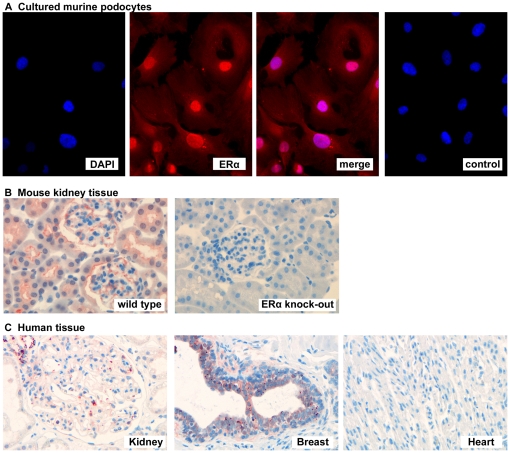
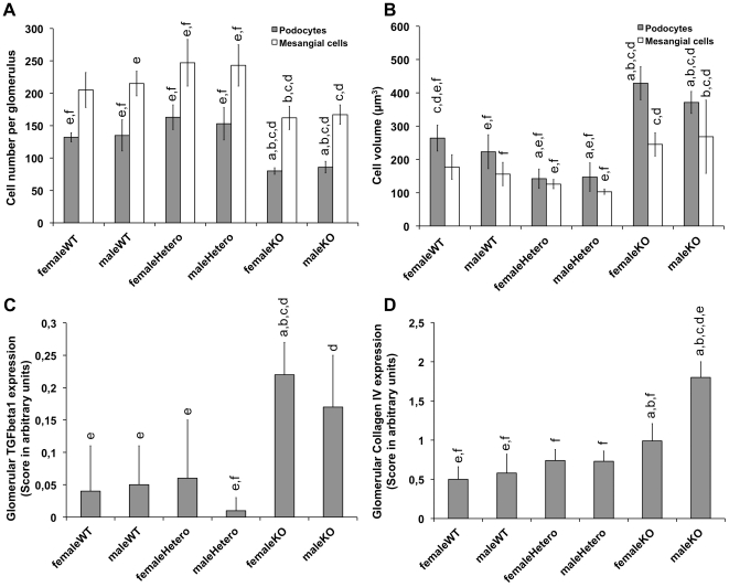
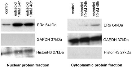
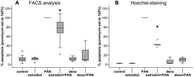
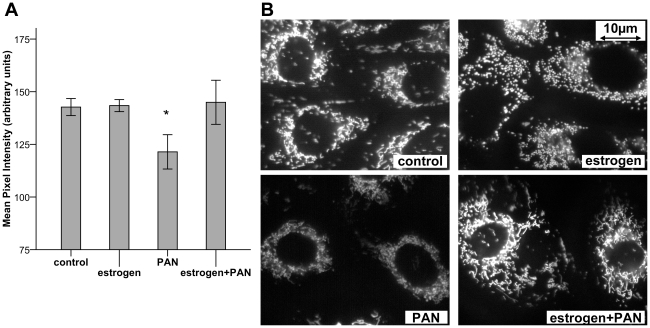
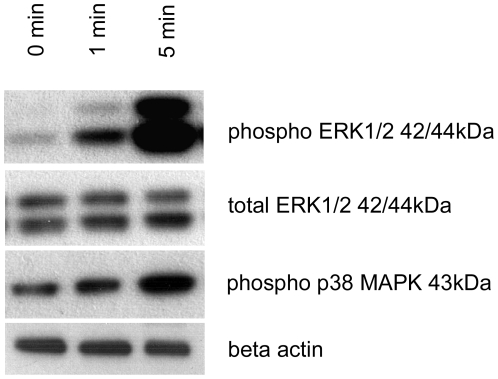
Expression of estrogen receptor alpha (ERα) was examined in podocytes in vitro and in vivo. Receptor localization was shown using Western blot of separated nuclear and cytoplasmatic protein fractions. Podocytes were treated with Puromycin aminonucleoside (PAN, apoptosis induction), estradiol, or both in combination. Apoptotic cells were detected with Hoechst nuclear staining and Annexin-FITC flow cytometry. To visualize mitochondrial membrane potential depolarization as an indicator for apoptosis, cells were stained with tetramethyl rhodamine methylester (TMRM). Estradiol-induced phosphorylation of ERK1/2 and p38 MAPK was examined by Western blot. Glomeruli of ERα knock-out mice and wild-type controls were analysed by histomorphometry and immunohistochemistry.
ERα was consistently expressed in human and murine podocytes. Estradiol stimulated ERα protein expression, reduced PAN-induced apoptosis in vitro by 26.5±24.6% or 56.6±5.9% (flow cytometry or Hoechst-staining, respectively; both p<0.05), and restored PAN-induced mitochondrial membrane potential depolarization. Estradiol enhanced ERK1/2 phosphorylation. In ERα knockout mice, podocyte number was reduced compared to controls (female/male: 80/86 vs. 132/135 podocytes per glomerulus, p<0.05). Podocyte volume was enhanced in ERα knockout mice (female/male: 429/371 µm(3) vs. 264/223 µm(3) in controls, p<0.05). Tgfβ1 and collagen type IV expression were increased in knockout mice, indicating glomerular damage.
Podocytes express ERα, whose activation leads to a significant protection against experimentally induced apoptosis. Possible underlying mechanisms include stabilization of mitochondrial membrane potential and activation of MAPK signalling. Characteristic morphological changes indicating glomerulopathy in ERα knock-out mice support the in vivo relevance of the ERα for podocyte viability and function. Thus, our findings provide a novel model for the protective influence of female gender on chronic glomerular diseases.
背景/目的:流行病学研究表明,女性在慢性肾脏疾病中的预后明显优于男性。这表明性别激素对肾小球结构和功能有重要影响。我们研究了雌二醇对足细胞的潜在直接保护作用。
在体外和体内检查足细胞中雌激素受体α(ERα)的表达。通过分离核和细胞质蛋白部分的 Western blot 显示受体定位。用嘌呤霉素氨基核苷(PAN,诱导凋亡)、雌二醇或两者联合处理足细胞。用 Hoechst 核染色和 Annexin-FITC 流式细胞术检测凋亡细胞。用四甲基罗丹明甲酯(TMRM)染色来可视化线粒体膜电位去极化作为凋亡的指标。通过 Western blot 检查雌二醇诱导的 ERK1/2 和 p38 MAPK 磷酸化。用组织形态计量学和免疫组织化学分析 ERα 敲除小鼠和野生型对照的肾小球。
ERα 在人源和鼠源足细胞中均有表达。雌二醇刺激 ERα 蛋白表达,体外减少 PAN 诱导的凋亡 26.5±24.6%或 56.6±5.9%(流式细胞术或 Hoechst 染色,均 p<0.05),并恢复 PAN 诱导的线粒体膜电位去极化。雌二醇增强 ERK1/2 磷酸化。在 ERα 敲除小鼠中,与对照组相比,足细胞数量减少(雌性/雄性:80/86 对 132/135 个足细胞/肾小球,p<0.05)。ERα 敲除小鼠中足细胞体积增大(雌性/雄性:429/371 µm³ 对 264/223 µm³ 在对照组中,p<0.05)。Tgfβ1 和胶原 IV 表达增加,表明肾小球损伤。
足细胞表达 ERα,其激活可显著对抗实验性诱导的凋亡。潜在的机制包括稳定线粒体膜电位和激活 MAPK 信号转导。ERα 敲除小鼠中特征性的形态学变化表明肾小球病,支持 ERα 对足细胞活力和功能的体内相关性。因此,我们的研究结果为女性性别对慢性肾小球疾病的保护作用提供了一个新的模型。