Department of Biochemistry and Molecular Biology, University of Nebraska Medical Center, Omaha, NE, USA.
Oncogene. 2012 Jul 12;31(28):3346-56. doi: 10.1038/onc.2011.505. Epub 2011 Nov 21.
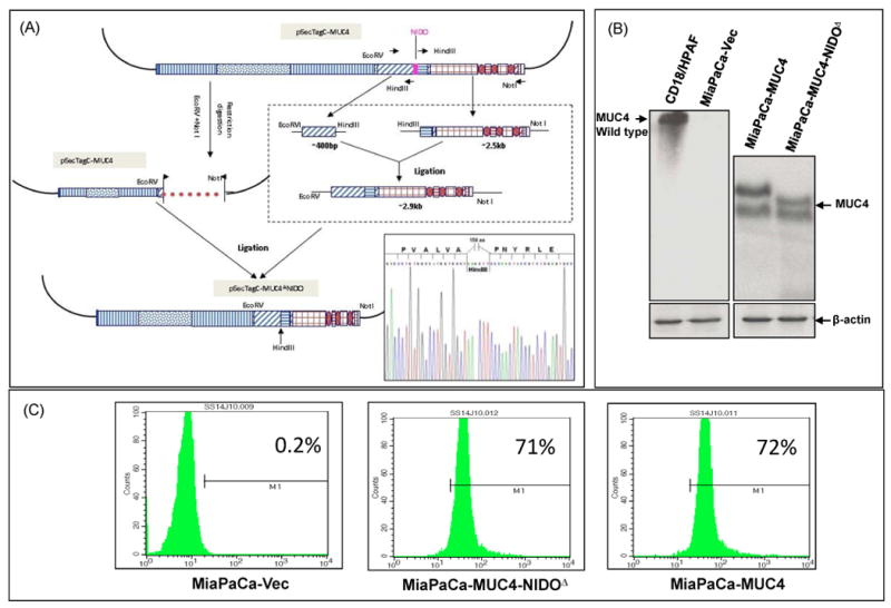
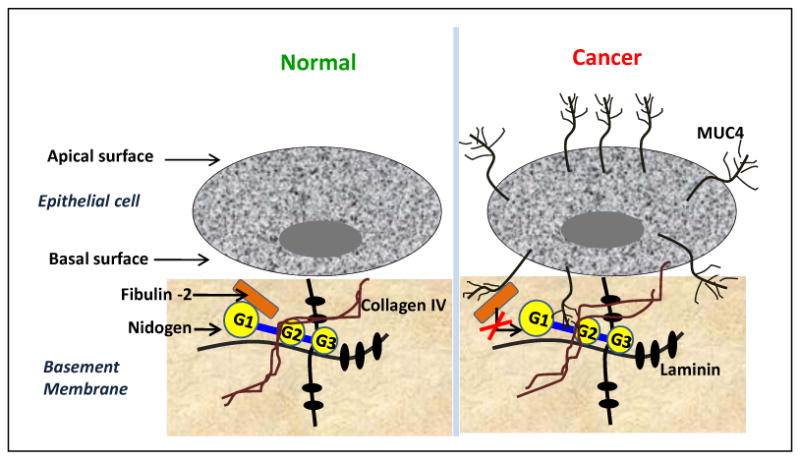
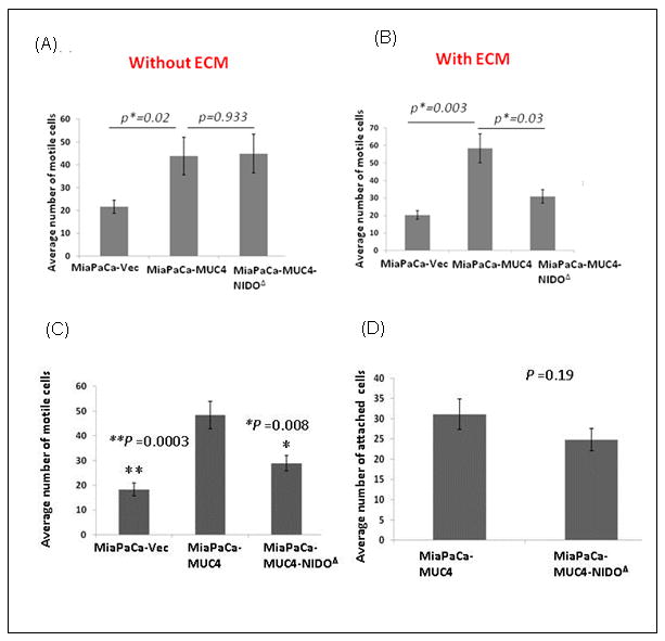
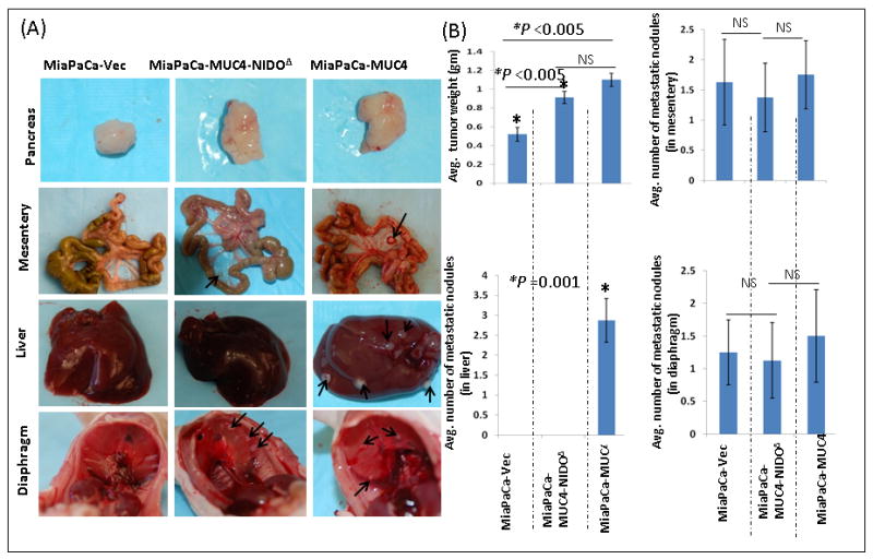
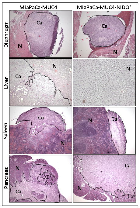
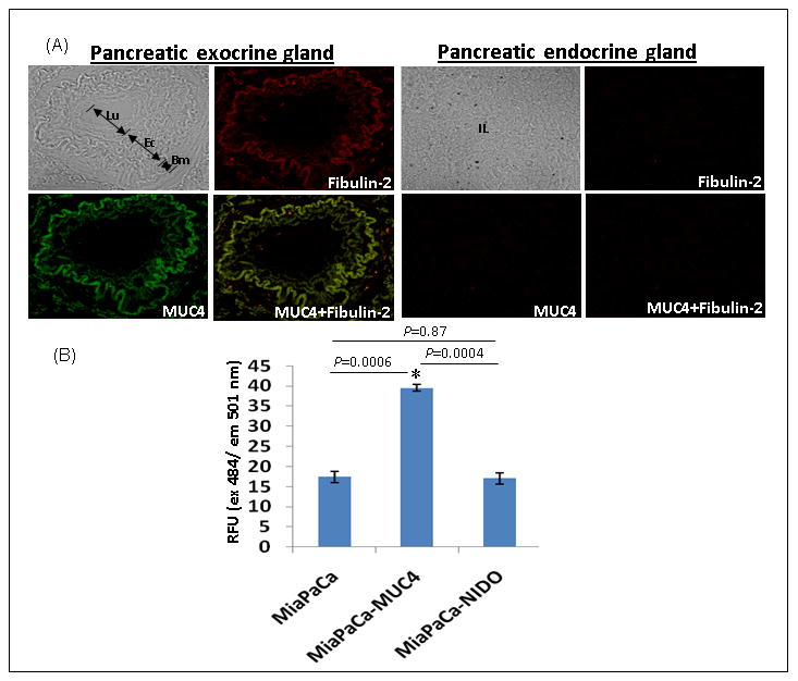
MUC4 is a large transmembrane type I glycoprotein that is overexpressed in pancreatic cancer (PC) and has been shown to be associated with its progression and metastasis. However, the exact cellular and molecular mechanism(s) through which MUC4 promotes metastasis of PC cells has been sparsely studied. Here we showed that the nidogen-like (NIDO) domain of MUC4, which is similar to the G1-domain present in the nidogen or entactin (an extracellular matrix protein), contributes to the protein-protein interaction property of MUC4. By this interaction, MUC4 promotes breaching of basement membrane (BM) integrity, and spreading of cancer cells. These observations are corroborated with the data from our study using an engineered MUC4 protein without the NIDO domain, which was ectopically expressed in the MiaPaCa PC cells, lacking endogenous MUC4 and nidogen protein. The in vitro studies demonstrated an enhanced invasiveness of MiaPaCa cells expressing MUC4 (MiaPaCa-MUC4) compared with vector-transfected cells (MiaPaCa-Vec; P=0.003) or cells expressing MUC4 without the NIDO domain (MiaPaCa-MUC4-NIDO(Δ); P=0.03). However, the absence of NIDO-domain has no significant role on cell growth and motility (P=0.93). In the in vivo studies, all the mice orthotopically implanted with MiPaCa-MUC4 cells developed metastasis to the liver as compared with MiaPaCa-Vec or the MiaPaCa-MUC4-NIDO(Δ) group, hence, supporting our in vitro observations. Additionally, a reduced binding (P=0.0004) of MiaPaCa-MUC4-NIDO(Δ) cells to the fibulin-2 coated plates compared with MiaPaCa-MUC4 cells indicated a possible interaction between the MUC4-NIDO domain and fibulin-2, a nidogen-interacting protein. Furthermore, in PC tissue samples, MUC4 colocalized with the fibulin-2 present in the BM. Altogether, our findings demonstrate that the MUC4-NIDO domain significantly contributes to the MUC4-mediated metastasis of PC cells. This may be partly due to the interaction between the MUC4-NIDO domain and fibulin-2.
黏蛋白 4(MUC4)是一种大型跨膜 I 型糖蛋白,在胰腺癌(PC)中过度表达,并且与肿瘤的进展和转移有关。然而,MUC4 促进 PC 细胞转移的确切细胞和分子机制尚未得到充分研究。在这里,我们发现 MUC4 的类巢蛋白(NIDO)结构域与巢蛋白或 entactin(一种细胞外基质蛋白)中的 G1 结构域相似,有助于 MUC4 的蛋白-蛋白相互作用特性。通过这种相互作用,MUC4 促进了基底膜(BM)完整性的破坏和癌细胞的扩散。这些观察结果与我们使用一种没有 NIDO 结构域的工程化 MUC4 蛋白进行的研究数据相吻合,该蛋白在缺乏内源性 MUC4 和巢蛋白的 MiaPaCa PC 细胞中异位表达。体外研究表明,与转染载体的细胞(MiaPaCa-Vec;P=0.003)或表达没有 NIDO 结构域的 MUC4 的细胞(MiaPaCa-MUC4-NIDO(Δ);P=0.03)相比,表达 MUC4 的 MiaPaCa 细胞(MiaPaCa-MUC4)的侵袭性增强。然而,NIDO 结构域的缺失对细胞生长和运动性没有显著影响(P=0.93)。在体内研究中,与 MiaPaCa-Vec 或 MiaPaCa-MUC4-NIDO(Δ)组相比,所有原位植入 MiPaCa-MUC4 细胞的小鼠均发展为肝转移,因此支持了我们的体外观察结果。此外,与 MiaPaCa-MUC4 细胞相比,MiaPaCa-MUC4-NIDO(Δ)细胞与纤连蛋白-2 包被的平板的结合减少(P=0.0004),表明 MUC4-NIDO 结构域和纤连蛋白-2 之间可能存在相互作用,纤连蛋白-2 是一种与巢蛋白相互作用的蛋白。此外,在 PC 组织样本中,MUC4 与 BM 中存在的纤连蛋白-2 共定位。总之,我们的研究结果表明,MUC4-NIDO 结构域显著促进了 PC 细胞的 MUC4 介导的转移。这可能部分是由于 MUC4-NIDO 结构域与纤连蛋白-2 之间的相互作用。