Department of Orthopaedics, University Medical Center Hamburg-Eppendorf, Germany.
Acta Orthop. 2011 Oct;82(5):596-601. doi: 10.3109/17453674.2011.625534.
Soft tissue necrobiosis and T-lymphocyte infiltration within the periprosthetic soft tissue have been linked to a suggested hypersensitivity reaction of the delayed-type following the metal-on-metal arthroplasty. While we observed both synovial necrobiosis and lymphocyte infiltrates in synovial tissues with failed metal-on-polyethylene prostheses, we hypothesized that both findings are unspecific for metal-on-metal bearing coupes. Thus, we wished to quantify the extent of necrobiosis and the amount of T-lymphocyte infiltration in aseptically loosened metal-on-polyethylene prostheses.
We analyzed 28 consecutive synovial biopsy specimens obtained at revision surgery of aseptically loosened metal-on-polyethylene prostheses (19 hips and 9 knees) and quantified both the extent of necrobiosis vertically and the density of CD3+, CD4+, and CD8+ lymphocytes within the joint capsular tissue. We excluded patients with inflammatory skeletal disease or with a history of metal hypersensitivity.
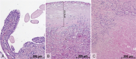
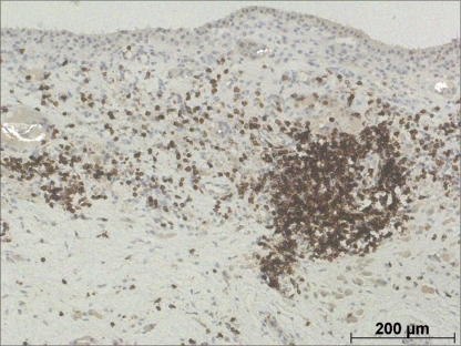
We found necrobiosis in 23 of 28 cases and it was most often connected with the superficial portions of the synovium. Necrobiosis of deeper tissues was seen in 8 specimens and it was strongly associated with superficial necrobiosis. While CD3+ lymphocytes were detected in each biopsy, 4 cases with more than 300 CD3+ lymphocytes were identified in the group of 26 cases that presented with more than 100 CD3+ lymphocytes within one high-power field. 16 cases with more than 100 CD3+ lymphocytes also showed concomitant superficial necrobiosis of the synovium. In the inflammatory infiltration of periprosthetic synovium, CD8+ lymphocytes predominated over CD4+ cells.
Synovial necrobiosis and infiltration of T-lymphocytes are common findings in tissues around aseptically loosened metal-on-polyethylene arthroplasty in patients without a clinically suspected metal hypersensitivity reaction. Thus, neither necrobiosis nor infiltration of T-lymphocytes should be considered to be specific for failed metal-on-metal bearings or metal hypersensitivity reaction.
软组织坏死和 T 淋巴细胞浸润在假体周围软组织中与金属对金属关节置换术后迟发型超敏反应有关。虽然我们在失败的金属对聚乙烯假体的滑膜组织中观察到滑膜坏死和淋巴细胞浸润,但我们假设这两种发现对金属对金属承窝均不具有特异性。因此,我们希望定量评估无菌性松动的金属对聚乙烯假体中坏死的程度和 T 淋巴细胞的浸润量。
我们分析了 28 例因无菌性松动的金属对聚乙烯假体翻修手术获得的连续滑膜活检标本(19 髋和 9 膝),并垂直定量测量了坏死的程度以及关节囊组织中 CD3+、CD4+和 CD8+T 淋巴细胞的密度。我们排除了患有炎症性骨病或有金属过敏史的患者。
我们发现 28 例中有 23 例存在坏死,且坏死最常与滑膜的浅层有关。8 例深部组织存在坏死,且与浅层坏死密切相关。虽然每个活检均检测到 CD3+淋巴细胞,但在 26 例中,有 4 例的 CD3+淋巴细胞超过 300 个/高倍视野,而这 26 例中有 4 例的 CD3+淋巴细胞超过 100 个/高倍视野。16 例 CD3+淋巴细胞超过 100 个/高倍视野的病例也伴有滑膜浅层坏死。在假体周围滑膜的炎症浸润中,CD8+淋巴细胞比 CD4+细胞更常见。
在无临床可疑金属过敏反应的无菌性松动的金属对聚乙烯关节置换术后,滑膜坏死和 T 淋巴细胞浸润是常见的组织学发现。因此,坏死和 T 淋巴细胞浸润都不应被认为是金属对金属假体失败或金属过敏反应的特异性表现。