• 文献检索
  • 文档翻译
  • 深度研究
  • 学术资讯
  • Suppr Zotero 插件Zotero 插件
  • 邀请有礼
  • 套餐&价格
  • 历史记录
应用&插件
Suppr Zotero 插件Zotero 插件浏览器插件Mac 客户端Windows 客户端微信小程序
定价
高级版会员购买积分包购买API积分包
服务
文献检索文档翻译深度研究API 文档MCP 服务
关于我们
关于 Suppr公司介绍联系我们用户协议隐私条款
关注我们

Suppr 超能文献

核心技术专利:CN118964589B侵权必究
粤ICP备2023148730 号-1Suppr @ 2026

文献检索

告别复杂PubMed语法,用中文像聊天一样搜索,搜遍4000万医学文献。AI智能推荐,让科研检索更轻松。

立即免费搜索

文件翻译

保留排版,准确专业,支持PDF/Word/PPT等文件格式,支持 12+语言互译。

免费翻译文档

深度研究

AI帮你快速写综述,25分钟生成高质量综述,智能提取关键信息,辅助科研写作。

立即免费体验

为什么放射摄影不应再被视为膝关节骨关节炎软骨纵向评估的替代结局指标。

Why radiography should no longer be considered a surrogate outcome measure for longitudinal assessment of cartilage in knee osteoarthritis.

机构信息

Quantitative Imaging Center, Department of Radiology, Boston University School of Medicine, 820 Harrison Avenue, FGH Building, 3rd Floor, Boston, MA 02118, USA.

出版信息

Arthritis Res Ther. 2011;13(6):247. doi: 10.1186/ar3488. Epub 2011 Nov 24.

DOI:10.1186/ar3488
PMID:22136179
原文链接:https://pmc.ncbi.nlm.nih.gov/articles/PMC3334624/
Abstract

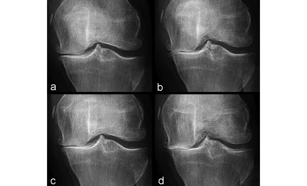
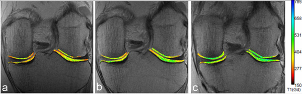
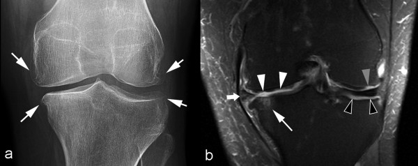
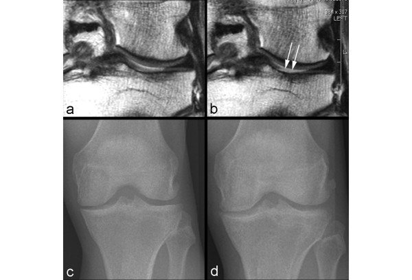
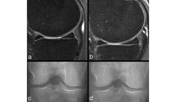
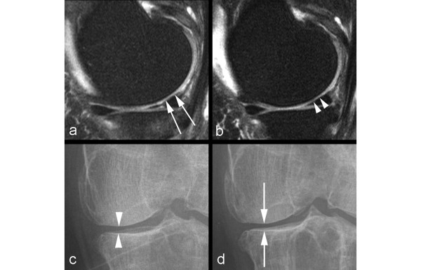
Imaging of cartilage has traditionally been achieved indirectly with conventional radiography. Loss of joint space width, or 'joint space narrowing', is considered a surrogate marker for cartilage thinning. However, radiography is severely limited by its inability to visualize cartilage, the difficulty of ascertaining the optimum and reproducible positioning of the joint in serial assessments, and the difficulty of grading joint space narrowing visually. With the availability of advanced magnetic resonance imaging (MRI) scanners, new pulse sequences, and imaging techniques, direct visualization of cartilage has become possible. MRI enables visualization not only of cartilage but also of other important features of osteoarthritis simultaneously. 'Pre-radiographic' cartilage changes depicted by MRI can be measured reliably by a semiquantitative or quantitative approach. MRI enables accurate measurement of longitudinal changes in quantitative cartilage morphology in knee osteoarthritis. Moreover, compositional MRI allows imaging of 'pre-morphologic' changes (that is, visualization of subtle intrasubstance matrix changes before any obvious morphologic alterations occur). Detection of joint space narrowing on radiography seems outdated now that it is possible to directly visualize morphologic and pre-morphologic changes of cartilage by using conventional as well as complex MRI techniques.

摘要

传统上,软骨的成像都是通过常规放射摄影术间接实现的。关节间隙变窄被认为是软骨变薄的替代标志物。然而,放射摄影术受到其无法可视化软骨、难以确定关节在连续评估中的最佳和可重复的定位以及难以直观地对关节间隙变窄进行分级等限制。随着先进磁共振成像(MRI)扫描仪、新脉冲序列和成像技术的出现,直接可视化软骨成为可能。MRI 不仅可以可视化软骨,还可以同时可视化骨关节炎的其他重要特征。MRI 可以通过半定量或定量方法可靠地测量“影像学前”软骨变化。MRI 可以准确测量膝关节骨关节炎定量软骨形态的纵向变化。此外,成分 MRI 允许对“前形态学”变化进行成像(即,在发生任何明显形态改变之前可视化细微的内部基质变化)。现在,通过使用常规和复杂的 MRI 技术,直接可视化软骨的形态学和前形态学变化,放射摄影术检测关节间隙变窄似乎已经过时。

https://cdn.ncbi.nlm.nih.gov/pmc/blobs/9f51/3334624/a0e14f8e6332/ar3488-6.jpg
https://cdn.ncbi.nlm.nih.gov/pmc/blobs/9f51/3334624/42c96e611d44/ar3488-1.jpg
https://cdn.ncbi.nlm.nih.gov/pmc/blobs/9f51/3334624/c191915c2c4c/ar3488-2.jpg
https://cdn.ncbi.nlm.nih.gov/pmc/blobs/9f51/3334624/6174f91b6be1/ar3488-3.jpg
https://cdn.ncbi.nlm.nih.gov/pmc/blobs/9f51/3334624/77f2f6105bc3/ar3488-4.jpg
https://cdn.ncbi.nlm.nih.gov/pmc/blobs/9f51/3334624/9c907bc7cd45/ar3488-5.jpg
https://cdn.ncbi.nlm.nih.gov/pmc/blobs/9f51/3334624/a0e14f8e6332/ar3488-6.jpg

相似文献

1
Why radiography should no longer be considered a surrogate outcome measure for longitudinal assessment of cartilage in knee osteoarthritis.为什么放射摄影不应再被视为膝关节骨关节炎软骨纵向评估的替代结局指标。
Arthritis Res Ther. 2011;13(6):247. doi: 10.1186/ar3488. Epub 2011 Nov 24.
2
Change in joint space width: hyaline articular cartilage loss or alteration in meniscus?关节间隙宽度的改变:透明关节软骨丢失还是半月板改变?
Arthritis Rheum. 2006 Aug;54(8):2488-95. doi: 10.1002/art.22016.
3
Contribution of regional 3D meniscus and cartilage morphometry by MRI to joint space width in fixed flexion knee radiography--a between-knee comparison in subjects with unilateral joint space narrowing.MRI 测量膝关节矢状面固定屈曲位时的半月板和软骨 3D 形态学参数与关节间隙狭窄的相关性:单侧膝关节间隙狭窄患者的双膝对比研究。
Eur J Radiol. 2013 Dec;82(12):e832-9. doi: 10.1016/j.ejrad.2013.08.041. Epub 2013 Sep 16.
4
No loss of cartilage volume over three years in patients with knee osteoarthritis as assessed by magnetic resonance imaging.通过磁共振成像评估,膝关节骨关节炎患者三年内软骨体积无损失。
Osteoarthritis Cartilage. 2002 Dec;10(12):929-37. doi: 10.1053/joca.2002.0849.
5
Magnetic resonance imaging (MRI) of articular cartilage in knee osteoarthritis (OA): morphological assessment.膝关节骨关节炎(OA)关节软骨的磁共振成像(MRI):形态学评估
Osteoarthritis Cartilage. 2006;14 Suppl A:A46-75. doi: 10.1016/j.joca.2006.02.026. Epub 2006 May 19.
6
Contribution of meniscal extrusion and cartilage loss to joint space narrowing in osteoarthritis.半月板挤出和软骨损伤对骨关节炎关节间隙变窄的作用。
Clin Radiol. 1999 Aug;54(8):502-6. doi: 10.1016/s0009-9260(99)90846-2.
7
Clinical, radiographic, molecular and MRI-based predictors of cartilage loss in knee osteoarthritis.膝关节骨关节炎软骨丢失的临床、影像学、分子和 MRI 预测因素。
Ann Rheum Dis. 2011 Jul;70(7):1223-30. doi: 10.1136/ard.2010.141382.
8
Hand joint space narrowing and osteophytes are associated with magnetic resonance imaging-defined knee cartilage thickness and radiographic knee osteoarthritis: data from the Osteoarthritis Initiative.手部关节间隙变窄和骨赘与磁共振成像定义的膝关节软骨厚度和放射学膝关节骨关节炎相关:来自骨关节炎倡议的数据。
J Rheumatol. 2012 Jan;39(1):161-6. doi: 10.3899/jrheum.110603. Epub 2011 Nov 1.
9
Comparison of Diagnostic Performance of Semi-Quantitative Knee Ultrasound and Knee Radiography with MRI: Oulu Knee Osteoarthritis Study.半定量膝关节超声与膝关节X线摄影对膝关节诊断性能的比较及与MRI的对比:奥卢膝关节骨关节炎研究
Sci Rep. 2016 Mar 1;6:22365. doi: 10.1038/srep22365.
10
Clinical and translational potential of MRI evaluation in knee osteoarthritis.MRI 评估在膝骨关节炎中的临床与转化潜力。
Curr Rheumatol Rep. 2014 Jan;16(1):391. doi: 10.1007/s11926-013-0391-6.

引用本文的文献

1
Development of a hip osteoarthritis index for gait quality assessment: a data-driven comparative study.用于步态质量评估的髋骨关节炎指数的开发:一项数据驱动的比较研究。
Sci Rep. 2025 Aug 5;15(1):28618. doi: 10.1038/s41598-025-10074-2.
2
Computational and Imaging Approaches for Precision Characterization of Bone, Cartilage, and Synovial Biomolecules.用于精确表征骨、软骨和滑膜生物分子的计算与成像方法
J Pers Med. 2025 Jul 9;15(7):298. doi: 10.3390/jpm15070298.
3
Clustering and classification of early knee osteoarthritis using machine-learning analysis of step-up and down test kinematics in recreational table tennis players.

本文引用的文献

1
Evolution of semi-quantitative whole joint assessment of knee OA: MOAKS (MRI Osteoarthritis Knee Score).膝关节骨关节炎半定量整体关节评估的演变:MOAKS(磁共振成像骨关节炎膝关节评分)。
Osteoarthritis Cartilage. 2011 Aug;19(8):990-1002. doi: 10.1016/j.joca.2011.05.004. Epub 2011 May 23.
2
Different thresholds for detecting osteophytes and joint space narrowing exist between the site investigators and the centralized reader in a multicenter knee osteoarthritis study--data from the Osteoarthritis Initiative.在一项多中心膝骨关节炎研究中,不同的中心研究者和中心化读片者之间存在骨赘和关节间隙狭窄检测的阈值差异——来自骨关节炎倡议的数据。
Skeletal Radiol. 2012 Feb;41(2):179-86. doi: 10.1007/s00256-011-1142-2. Epub 2011 Apr 9.
3
利用娱乐乒乓球运动员上下台阶测试运动学的机器学习分析对早期膝关节骨关节炎进行聚类和分类
PeerJ. 2025 May 27;13:e19471. doi: 10.7717/peerj.19471. eCollection 2025.
4
ShapeMed-Knee: A Dataset and Neural Shape Model Benchmark for Modeling 3D Femurs.ShapeMed-膝关节:用于3D股骨建模的数据集和神经形状模型基准
IEEE Trans Med Imaging. 2025 Mar;44(3):1140-1152. doi: 10.1109/TMI.2024.3485613. Epub 2025 Mar 17.
5
Assessment of validity, reliability, and feasibility of OMERACT ultrasound knee osteoarthritis scores in Egyptian patients with primary knee osteoarthritis.评估 OMERACT 膝关节骨关节炎评分在埃及原发性膝关节骨关节炎患者中的有效性、可靠性和可行性。
Clin Rheumatol. 2024 Dec;43(12):3913-3923. doi: 10.1007/s10067-024-07171-4. Epub 2024 Oct 18.
6
Comparative evaluation of knee osteoarthritis: radiography vs. ultrasonography in Nepalese population: a cross-sectional study.尼泊尔人群膝关节骨关节炎的比较评估:X线摄影与超声检查对比:一项横断面研究
Ann Med Surg (Lond). 2024 Jul 22;86(9):5131-5136. doi: 10.1097/MS9.0000000000002393. eCollection 2024 Sep.
7
Investigating the relationship between radiographic joint space width loss and deep learning-derived magnetic resonance imaging-based cartilage thickness loss in the medial weight-bearing region of the tibiofemoral joint.研究胫股关节内侧负重区域X线片关节间隙宽度损失与基于深度学习的磁共振成像软骨厚度损失之间的关系。
Osteoarthr Cartil Open. 2024 Aug 3;6(3):100508. doi: 10.1016/j.ocarto.2024.100508. eCollection 2024 Sep.
8
Single Stance Radiography of the Knee Joint - A Novel Approach to Assess the Degree of Knee Osteoarthritis.膝关节单腿站立位X线摄影——一种评估膝关节骨关节炎程度的新方法。
J Orthop Case Rep. 2024 May;14(5):184-189. doi: 10.13107/jocr.2024.v14.i05.4476.
9
ShapeMed-Knee: A Dataset and Neural Shape Model Benchmark for Modeling 3D Femurs.ShapeMed-Knee:用于3D股骨建模的数据集和神经形状模型基准。
medRxiv. 2024 Oct 22:2024.05.06.24306965. doi: 10.1101/2024.05.06.24306965.
10
Approaches to optimize analyses of multidimensional ordinal MRI data in osteoarthritis research: A perspective.骨关节炎研究中优化多维有序MRI数据分析的方法:一种观点。
Osteoarthr Cartil Open. 2024 Mar 27;6(2):100465. doi: 10.1016/j.ocarto.2024.100465. eCollection 2024 Jun.
Summary and recommendations of the OARSI FDA osteoarthritis Assessment of Structural Change Working Group.
OARSI FDA 骨关节炎结构改变评估工作组的总结和建议。
Osteoarthritis Cartilage. 2011 May;19(5):606-10. doi: 10.1016/j.joca.2011.02.018. Epub 2011 Mar 23.
4
Responsiveness and reliability of MRI in knee osteoarthritis: a meta-analysis of published evidence.MRI 在膝骨关节炎中的反应性和可靠性:已发表证据的荟萃分析。
Osteoarthritis Cartilage. 2011 May;19(5):589-605. doi: 10.1016/j.joca.2010.10.030. Epub 2011 Mar 23.
5
Systematic review of the concurrent and predictive validity of MRI biomarkers in OA.MRI 生物标志物在骨关节炎中的并发和预测有效性的系统评价。
Osteoarthritis Cartilage. 2011 May;19(5):557-88. doi: 10.1016/j.joca.2010.10.029. Epub 2011 Mar 23.
6
Characterization of human osteoarthritic cartilage using optical and magnetic resonance imaging.使用光学和磁共振成像技术对人骨性关节炎软骨进行特征描述。
Mol Imaging Biol. 2012 Feb;14(1):32-9. doi: 10.1007/s11307-011-0480-8.
7
MRI-based extended ordered values more efficiently differentiate cartilage loss in knees with and without joint space narrowing than region-specific approaches using MRI or radiography--data from the OA initiative.基于 MRI 的扩展有序值比使用 MRI 或放射摄影的基于特定区域的方法更有效地区分有无关节间隙变窄的膝关节软骨丢失——来自 OA 倡议的数据。
Osteoarthritis Cartilage. 2011 Jun;19(6):689-99. doi: 10.1016/j.joca.2011.02.011. Epub 2011 Feb 19.
8
Osteoarthritis year 2010 in review: imaging.骨关节炎 2010 年年鉴:影像学。
Osteoarthritis Cartilage. 2011 Apr;19(4):354-60. doi: 10.1016/j.joca.2011.02.003. Epub 2011 Feb 12.
9
Imaging in osteoarthritis: what have we learned and where are we going?骨关节炎的影像学:我们已经学到了什么,我们的未来方向在哪里?
Minerva Med. 2011 Feb;102(1):15-32.
10
Articular cartilage in the knee: current MR imaging techniques and applications in clinical practice and research.膝关节软骨:当前的磁共振成像技术及其在临床实践和研究中的应用。
Radiographics. 2011 Jan-Feb;31(1):37-61. doi: 10.1148/rg.311105084.