Department of Urology, Hospital de São João, Alameda Professor Hernâni Monteiro, 4200-319, Porto-Portugal.
BMC Urol. 2012 Jan 4;12:1. doi: 10.1186/1471-2490-12-1.
Onabotulinumtoxin A (OnabotA) injection has been investigated as a novel treatment for benign prostatic enlargement caused by benign prostatic hyperplasia. An OnabotA-induced volume reduction caused by sympathetic fibers impairment has been proposed as a potential mechanism of action. Our aim was to investigate the expression of apoptosis-regulating proteins in the rat prostate following OnabotA intraprostatic injection.
Adult Wistar rats were injected in the ventral lobes of the prostate with 10 U of OnabotA or saline. A set of OnabotA-injected animals was further treated with 0.5 mg/kg of phenylephrine (PHE) subcutaneously daily. All animals were sacrificed after 1 week and had their prostates harvested. Immunohistochemical staining was performed for Bax, Bcl-xL and caspase-3 proteins and visualized by the avidin-biotin method. The optical density of the glandular cells was also determined, with measurement of differences between average optical densities for each group.
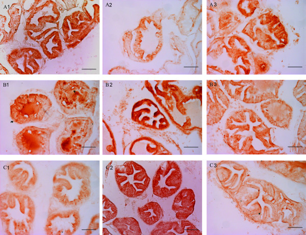
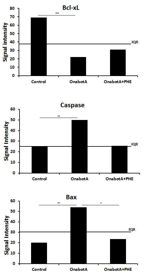
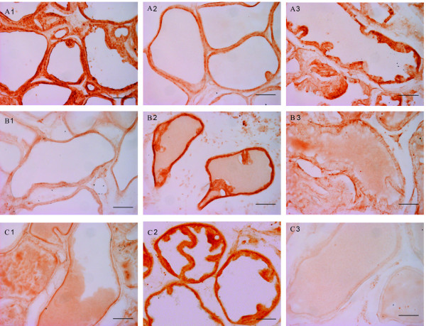
Saline-treated animals showed intense epithelial staining for Bcl-xL and a faint labelling for both Bax and Caspase-3. OnabotA-treated rats showed a reduced epithelial staining of Bcl-xL and a consistently increased Bax and Caspase-3 staining when compared with saline-treated animals. PHE-treated animals showed a stronger Bcl-xL staining and reduced staining of both Bax and Caspase-3 when compared to the OnabotA group. Mean signal intensity measurements for each immunoreaction confirmed a significant decrease of the signal intensity for Bcl-xL and a significant increase of the signal intensity for Bax and Caspase 3 in OnabotA-injected animals when compared with the control group. In OnabotA+PHE treated animals mean signal intensity for Bcl-xL, Bax and Caspase 3 immunoreactions was identical to that of the control animals.
These results support the hypothesis that OnabotA activates apoptotic pathways in the rat prostate through a mechanism that involves sympathetic outflow impairment.
肉毒杆菌毒素 A(OnabotA)注射已被研究作为一种治疗良性前列腺增生引起的良性前列腺增生的新方法。一种认为 OnabotA 通过损伤交感神经纤维引起的体积减少是其潜在的作用机制。我们的目的是研究 OnabotA 前列腺内注射后大鼠前列腺中凋亡调节蛋白的表达。
成年 Wistar 大鼠在前列腺腹叶注射 10U 的 OnabotA 或生理盐水。一组 OnabotA 注射动物进一步每天皮下给予 0.5mg/kg 苯肾上腺素(PHE)治疗。所有动物在 1 周后处死,采集前列腺。用抗生物素蛋白法进行 Bax、Bcl-xL 和 caspase-3 蛋白的免疫组织化学染色,并进行可视化。还测定了腺细胞的光密度,测量了每组平均光密度的差异。
生理盐水处理的动物显示 Bcl-xL 上皮染色强烈,Bax 和 Caspase-3 染色微弱。与生理盐水处理的动物相比,OnabotA 处理的大鼠显示 Bcl-xL 上皮染色减少,Bax 和 Caspase-3 染色增加。与 OnabotA 组相比,PHE 处理的动物显示出更强的 Bcl-xL 染色和 Bax 和 Caspase-3 染色减少。每种免疫反应的平均信号强度测量证实,与对照组相比,OnabotA 注射动物的 Bcl-xL 信号强度显著降低,Bax 和 Caspase 3 的信号强度显著增加。在 OnabotA+PHE 处理的动物中,Bcl-xL、Bax 和 Caspase 3 免疫反应的平均信号强度与对照动物相同。
这些结果支持了这样的假设,即 OnabotA 通过涉及交感神经输出损伤的机制激活大鼠前列腺中的凋亡途径。