Key Laboratory for NeuroInformation of Ministry of Education, School of Life Science and Technology, University of Electronic Science and Technology of China, Chengdu, China.
PLoS One. 2011;7(1):e28196. doi: 10.1371/journal.pone.0028196. Epub 2012 Jan 5.
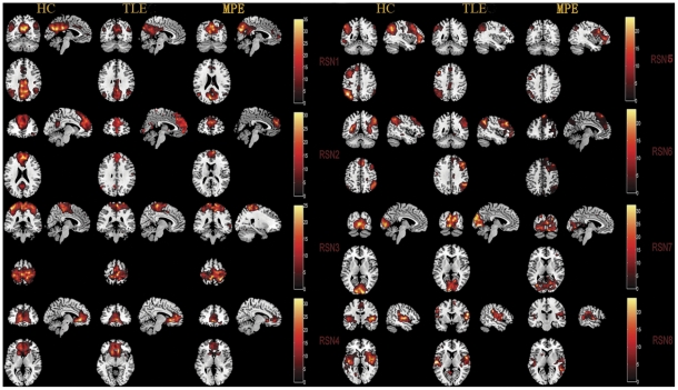
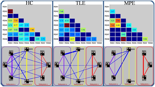
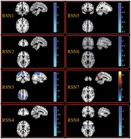
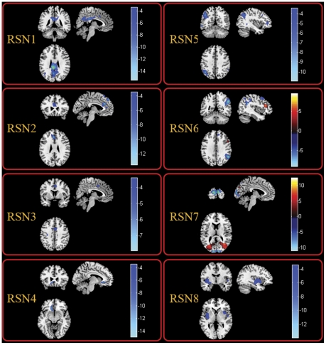
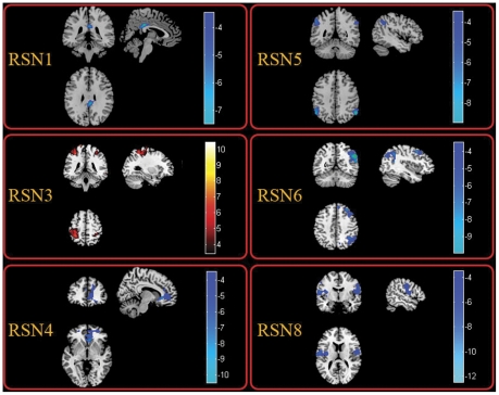
Examining the spontaneous activity to understand the neural mechanism of brain disorder is a focus in recent resting-state fMRI. In the current study, to investigate the alteration of brain functional connectivity in partial epilepsy in a systematical way, two levels of analyses (functional connectivity analysis within resting state networks (RSNs) and functional network connectivity (FNC) analysis) were carried out on resting-state fMRI data acquired from the 30 participants including 14 healthy controls(HC) and 16 partial epilepsy patients. According to the etiology, all patients are subdivided into temporal lobe epilepsy group (TLE, included 7 patients) and mixed partial epilepsy group (MPE, 9 patients). Using group independent component analysis, eight RSNs were identified, and selected to evaluate functional connectivity and FNC between groups. Compared with the controls, decreased functional connectivity within all RSNs was found in both TLE and MPE. However, dissociating patterns were observed within the 8 RSNs between two patient groups, i.e, compared with TLE, we found decreased functional connectivity in 5 RSNs increased functional connectivity in 1 RSN, and no difference in the other 2 RSNs in MPE. Furthermore, the hierarchical disconnections of FNC was found in two patient groups, in which the intra-system connections were preserved for all three subsystems while the lost connections were confined to intersystem connections in patients with partial epilepsy. These findings may suggest that decreased resting state functional connectivity and disconnection of FNC are two remarkable characteristics of partial epilepsy. The selective impairment of FNC implicated that it is unsuitable to understand the partial epilepsy only from global or local perspective. We presumed that studying epilepsy in the multi-perspective based on RSNs may be a valuable means to assess the functional changes corresponding to specific RSN and may contribute to the understanding of the neuro-pathophysiological mechanism of epilepsy.
研究自发活动以了解大脑障碍的神经机制是近期静息态 fMRI 的一个重点。在本研究中,为了系统地研究部分性癫痫中的脑功能连接改变,我们对来自 30 名参与者(包括 14 名健康对照者[HC]和 16 名部分性癫痫患者)的静息态 fMRI 数据进行了两个层次的分析(静息态网络内的功能连接分析[RSNs]和功能网络连接[FNC]分析)。根据病因,所有患者均进一步分为颞叶癫痫组(TLE,包括 7 名患者)和混合性部分性癫痫组(MPE,9 名患者)。使用组独立成分分析,我们确定了 8 个 RSN,并选择其来评估组间功能连接和 FNC。与对照组相比,TLE 和 MPE 患者的所有 RSN 内的功能连接均降低。然而,在两组患者之间的 8 个 RSN 中观察到了分离模式,即与 TLE 相比,我们发现 MPE 患者的 5 个 RSN 内的功能连接降低,1 个 RSN 内的功能连接增加,而另外 2 个 RSN 内的功能连接没有差异。此外,我们在两组患者中发现了 FNC 的层次脱连接,其中系统内连接在所有三个子系统中均保留,而部分性癫痫患者的连接丢失仅限于系统间连接。这些发现可能表明,静息态功能连接的降低和 FNC 的脱连接是部分性癫痫的两个显著特征。FNC 的选择性损伤表明,仅从全局或局部角度理解部分性癫痫是不合适的。我们推测,基于 RSN 从多视角研究癫痫可能是评估与特定 RSN 对应的功能变化的一种有价值的手段,并有助于理解癫痫的神经病理生理机制。