Miyata Kaori, Sukata Tokuo, Kushida Masahiko, Ogata Keiko, Suzuki Manabu, Ozaki Masakazu, Ozaki Keisuke, Uwagawa Satoshi
J Toxicol Pathol. 2009 Sep;22(3):199-203. doi: 10.1293/tox.22.199. Epub 2009 Oct 15.
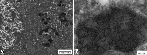
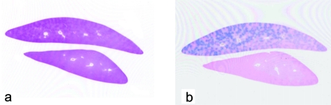
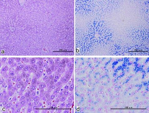
Spontaneous iron accumulation in hepatocytes was observed in a 7-week-old female Han Wistar GALAS rat. Very fine yellowish brown pigments, which showed a positive reaction with Berlin Blue stain, were apparent in the cytoplasm close to the bile canaliculi, with a diminishing periportal-to-centrilobular gradient. There were also differences in distribution between and within lobes. Transmission electron microscopy revealed cytosolic ferritin and pericanalicular siderosomes in hepatocytes. No degeneration or necrotic changes were observed, and non-hepatocyte cells did not demonstrate any obvious accumulation of iron. There were no abnormalities in the animal other than this finding in the liver.
在一只7周龄的雌性韩系Wistar GALAS大鼠中观察到肝细胞内的自发性铁蓄积。在靠近胆小管的细胞质中可见非常细小的黄褐色色素,其对柏林蓝染色呈阳性反应,门周至中央静脉的梯度逐渐减小。叶间和叶内的分布也存在差异。透射电子显微镜显示肝细胞中有胞质铁蛋白和胆小管周围的含铁小体。未观察到变性或坏死变化,非肝细胞也未显示出明显的铁蓄积。除肝脏的这一发现外,该动物无其他异常。