Department of Neuroscience, University of Uppsala, Uppsala, Sweden.
Int J Obes (Lond). 2013 Feb;37(2):230-6. doi: 10.1038/ijo.2012.13. Epub 2012 Jan 31.
Obesity adversely affects frontal lobe brain structure and function. Here we sought to show that people who are obese versus those who are of normal weight over a 5-year period have differential global and regional brain volumes.
Using voxel-based morphometry, contrasts were done between those who were recorded as being either obese or of normal weight over two time points in the 5 years prior to the brain scan. In a post-hoc preliminary analysis, we compared scores for obese and normal weight people who completed the trail-making task.
A total of 292 subjects were examined following exclusions (for example, owing to dementia, stroke and cortical infarcts) from the Prospective Investigation of the Vasculature in Uppsala Seniors cohort with a body mass index of normal weight (<25 kg m(-2)) or obese (30 kg m(-2)).
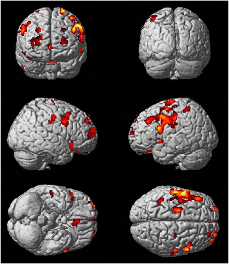
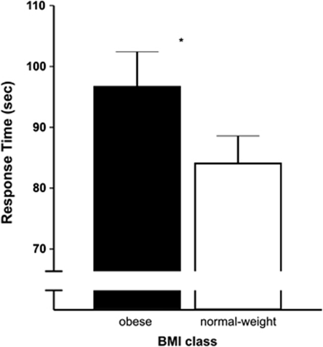
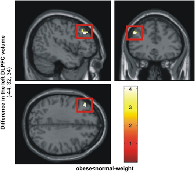
People who were obese had significantly smaller total brain volumes and specifically, significantly reduced total gray matter (GM) volume (GMV) (with no difference in white matter or cerebrospinal fluid). Initial exploratory whole brain uncorrected analysis revealed that people who were obese had significantly smaller GMV in the bilateral supplementary motor area, bilateral dorsolateral prefrontal cortex (DLPFC), left inferior frontal gyrus and left postcentral gyrus. Secondary more stringent corrected analyses revealed a surviving cluster of GMV difference in the left DLPFC. Finally, post-hoc contrasts of scores on the trail-making task, which is linked to DLPFC function, revealed that obese people were significantly slower than those of normal weight.
These findings suggest that in comparison with normal weight, people who are obese have smaller GMV, particularly in the left DLPFC. Our results may provide evidence for a potential working memory mechanism for the cognitive suppression of appetite that may lower the risk of developing obesity in later life.
肥胖会对额叶脑结构和功能产生不良影响。本研究旨在证明,在进行脑部扫描前的 5 年内,体重持续处于肥胖状态的人与体重正常的人相比,其大脑的整体和区域体积存在差异。
采用基于体素的形态测量学方法,对在 5 年内的两次脑部扫描前,分别被记录为肥胖或体重正常的人进行对比。在一项事后初步分析中,我们比较了完成连线测试任务的肥胖者和体重正常者的分数。
在排除了痴呆、中风和皮质梗死等情况后,对 Prospective Investigation of the Vasculature in Uppsala Seniors 队列中的 292 名受试者进行了检查,这些受试者的体质指数(BMI)正常(<25kg/m²)或肥胖(≥30kg/m²)。
肥胖者的总脑容量明显较小,特别是总灰质(GM)体积明显减少(WM 和脑脊液无差异)。初始的全脑未校正分析显示,肥胖者的双侧辅助运动区、双侧背外侧前额叶皮质(DLPFC)、左侧额下回和左侧后中央回的 GM 体积明显较小。经过更严格的校正分析,发现左侧 DLPFC 的 GM 体积差异仍存在一个存活的聚类。最后,对连线测试任务(与 DLPFC 功能相关)的分数进行事后对比发现,肥胖者的得分明显低于体重正常者。
与体重正常者相比,肥胖者的 GM 体积较小,特别是在左侧 DLPFC。我们的研究结果可能为食欲认知抑制的潜在工作记忆机制提供了证据,这种机制可能降低晚年肥胖的风险。